Copyright
©The Author(s) 2017.
World J Orthop. Oct 18, 2017; 8(10): 809-813
Published online Oct 18, 2017. doi: 10.5312/wjo.v8.i10.809
Published online Oct 18, 2017. doi: 10.5312/wjo.v8.i10.809
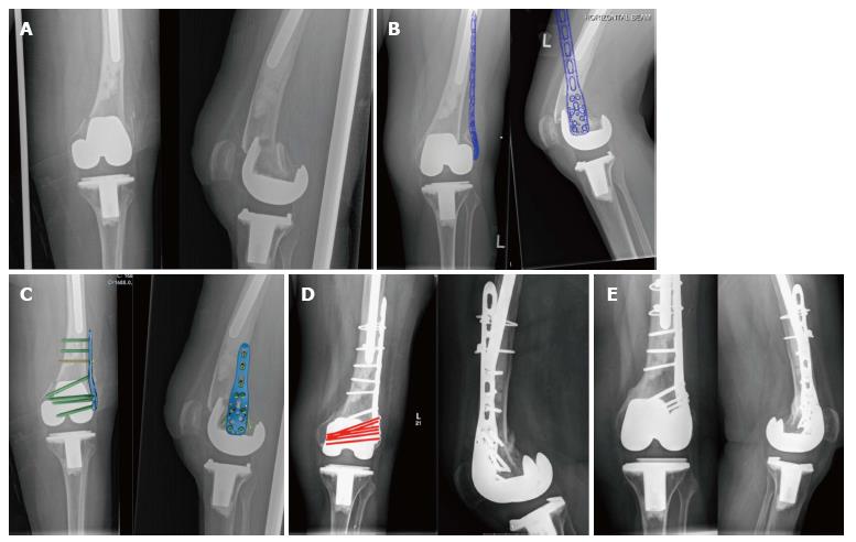
Figure 1 Radiograph series of patient A.
A: Anteroposterior and lateral injury radiographs; B: Templated anteroposterior radiograph showing proposed position of implant; C: Anteroposterior and lateral radiographs with PHILOS plate image superimposed to show orientation of screws; D: Post-operative radiographs at 6 mo with orientation of screws behind the femoral component shown in red; E: Anteroposterior and lateral radiographs at 19 mo post-op.
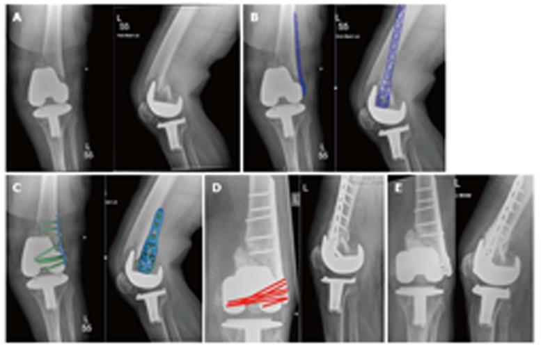
Figure 2 Radiograph series of patient B.
A: Anteroposterior and lateral injury radiographs; B: Templated anteroposterior radiograph showing proposed position of implant; C: Anteroposterior and lateral radiographs with PHILOS plate image superimposed to show orientation of screws; D: Post-operative radiographs at 5 mo with orientation of screws behind the femoral component shown in red; E: Anteroposterior and lateral radiographs at 16 mo post-op.
- Citation: Donnelly KJ, Tucker A, Ruiz A, Thompson NW. Managing extremely distal periprosthetic femoral supracondylar fractures of total knee replacements - a new PHILOS-ophy. World J Orthop 2017; 8(10): 809-813
- URL: https://www.wjgnet.com/2218-5836/full/v8/i10/809.htm
- DOI: https://dx.doi.org/10.5312/wjo.v8.i10.809


