Copyright
©The Author(s) 2015.
World J Orthop. Jan 18, 2015; 6(1): 127-136
Published online Jan 18, 2015. doi: 10.5312/wjo.v6.i1.127
Published online Jan 18, 2015. doi: 10.5312/wjo.v6.i1.127
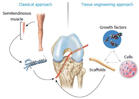
Figure 1 Comparison of the current clinical strategy in anterior cruciate ligament surgery to tissue engineering approaches.
The current “golden standard” in the clinical routine is the use of autologous tissue grafts such as semitendinosus (depicted in the figure) or patellar tendon. In tissue engineering approaches, scaffolds alone or in a combined fashion with cells or growth factors are used to improve tissue regeneration.
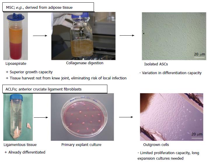
Figure 2 Overview of the main cell types used for anterior cruciate ligament tissue engineering approaches.
Two different types of cells are mainly regarded as the primary choice for anterior cruciate ligament (ACL) regeneration: mesenchymal stem cells (MSC) and ACL fibroblasts. Since MSCs can be isolated from adipose tissue (in our studies in cooperation with the Red Cross Blood Transfusion Service of Upper Austria, Linz, Austria) or bone aspirates, their harvest is less delicate than cells isolated from ligamentous tissue. Further advantages of MSCs over ACL fibroblasts are their superior growth capacity and capability of differentiating into the appropriate cell types. Nevertheless, due to their origin, ACL fibroblasts would be the accurate cell type to build up neoligamentous tissue. ASCs: Adipose derived stem cells.
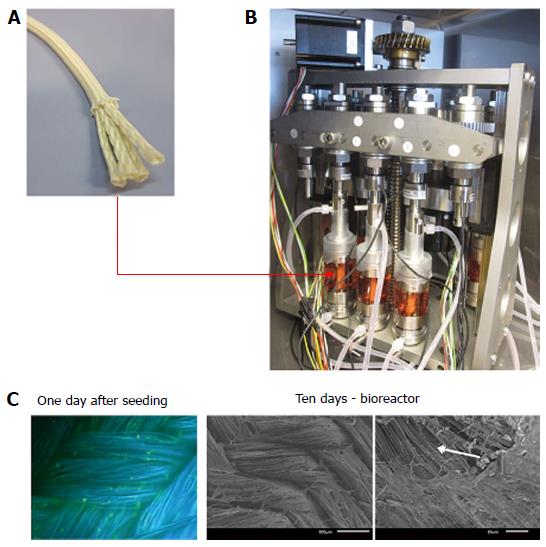
Figure 3 Adipose-derived stem cells cultured on silk-based ligament grafts (A) produce sheets of extracellular matrix proteins (C) under mechanical stimulation via a custom-made bioreactor system (B: design and construction in cooperation with the Technical University of Vienna, Institute of Materials Science and Technology).
A: The silk-based anterior cruciate ligament (ACL) scaffold is produced of Bombyx mori silk fibers in a wire-rope design; B: The scaffold is seeded with ASCs for 24 h and then transferred into bioreactor and cultured under linear and rotational displacement for 10 d. The mechanically stimulated ACL scaffolds show sheets of extracellular matrix. The arrow in the bottom panel indicates an artefact of scanning electron microscopy preparation. In this area, the covering extracellular matrix sheet has been flushed away due to too intense flushing, allowing the view to the underlying silk fibers.
- Citation: Nau T, Teuschl A. Regeneration of the anterior cruciate ligament: Current strategies in tissue engineering. World J Orthop 2015; 6(1): 127-136
- URL: https://www.wjgnet.com/2218-5836/full/v6/i1/127.htm
- DOI: https://dx.doi.org/10.5312/wjo.v6.i1.127


