Copyright
©2014 Baishideng Publishing Group Inc.
World J Orthop. Nov 18, 2014; 5(5): 566-573
Published online Nov 18, 2014. doi: 10.5312/wjo.v5.i5.566
Published online Nov 18, 2014. doi: 10.5312/wjo.v5.i5.566
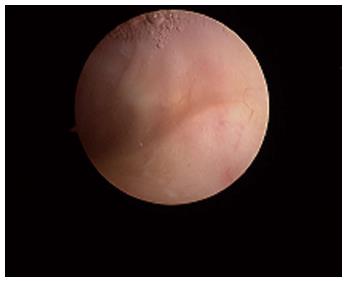
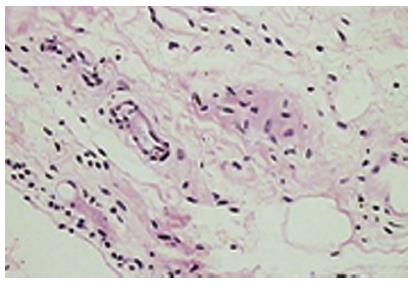
Figure 1 The normal synovium.
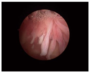
Figure 2 The synovium in rheumatoid arthritis.
Note the villous appearance and straight vessels.
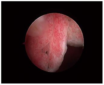
Figure 3 The synovium in psoriatic arthritis.
Note the hypervascular villous hypertrophy and tortuous vessels.
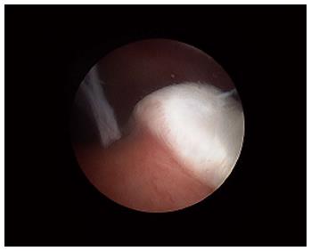
Figure 4 The cartilage-pannus junction.
Figure 5 The normal (areolar) synovium.
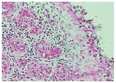
Figure 6 Lymphoid follicles in rheumatoid arthritis.
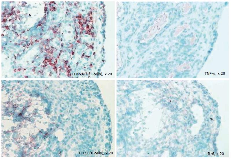
Figure 7 Differential staining of synovial biopsy for T cells, tumour necrosis factor, B cells (CD22), and interleukin-6 in a patient with active rheumatoid arthritis prior to disease-modifying therapy.
TNF: Tumour necrosis factor; IL: Interleukin.
- Citation: Wechalekar MD, Smith MD. Utility of arthroscopic guided synovial biopsy in understanding synovial tissue pathology in health and disease states. World J Orthop 2014; 5(5): 566-573
- URL: https://www.wjgnet.com/2218-5836/full/v5/i5/566.htm
- DOI: https://dx.doi.org/10.5312/wjo.v5.i5.566


