Copyright
©2014 Baishideng Publishing Group Inc.
World J Orthop. Sep 18, 2014; 5(4): 444-449
Published online Sep 18, 2014. doi: 10.5312/wjo.v5.i4.444
Published online Sep 18, 2014. doi: 10.5312/wjo.v5.i4.444
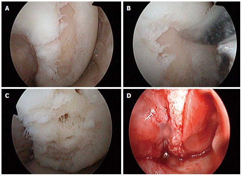
Figure 1 Twenty years old female with a chondral defect on the lateral condyl after trauma.
The AMIC® technique was done arthroscopic assisted: after debridement of the chondral defect (A), numerous perforations of the subchondral lamina were performed (B, C). The implantation of the matrix was performed under dry, arthroscopic conditions, as published before (D)[27].
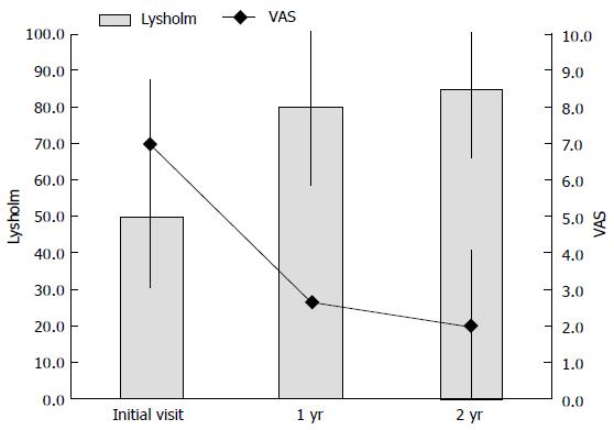
Figure 2 Significant improvements of the mean Lysholm and visual analogue scale score after 1 year and further increased values up to 2 years postoperatively in patients with cartilage knee defects treated with AMIC®[32].
VAS: Visual analogue scale.
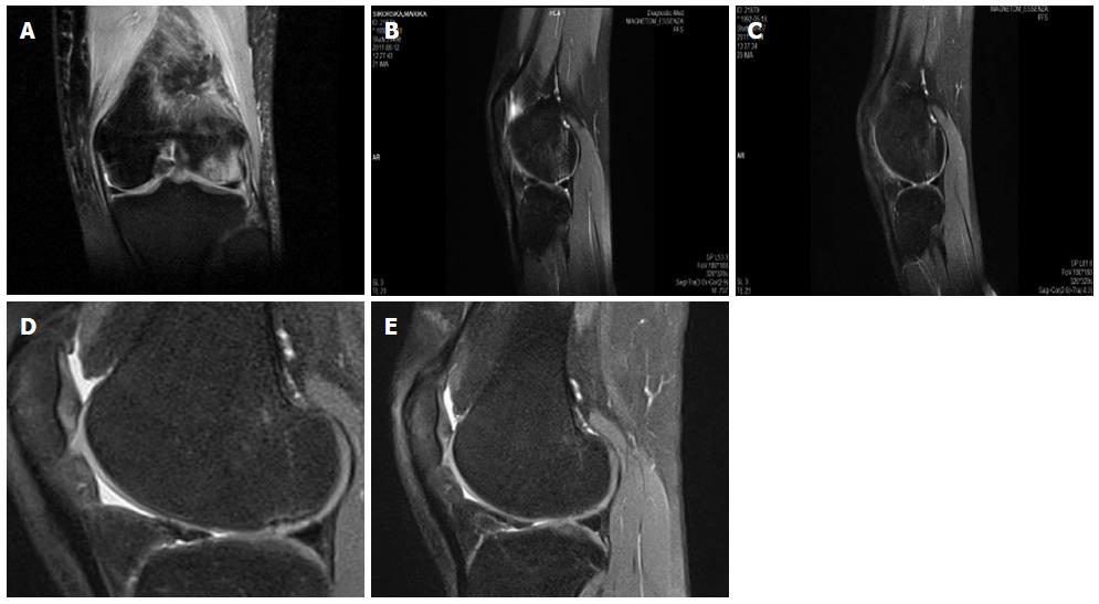
Figure 3 The same patient showing enhanced defect filling demonstrated by follow-up magnetic resonance imaging before surgery (A) and 3 (B), 6 (C), 12 (D) and 24 mo (E) after the index procedure.
- Citation: Bark S, Piontek T, Behrens P, Mkalaluh S, Varoga D, Gille J. Enhanced microfracture techniques in cartilage knee surgery: Fact or fiction? World J Orthop 2014; 5(4): 444-449
- URL: https://www.wjgnet.com/2218-5836/full/v5/i4/444.htm
- DOI: https://dx.doi.org/10.5312/wjo.v5.i4.444


