Copyright
©2013 Baishideng Publishing Group Co.
World J Orthop. Oct 18, 2013; 4(4): 327-332
Published online Oct 18, 2013. doi: 10.5312/wjo.v4.i4.327
Published online Oct 18, 2013. doi: 10.5312/wjo.v4.i4.327
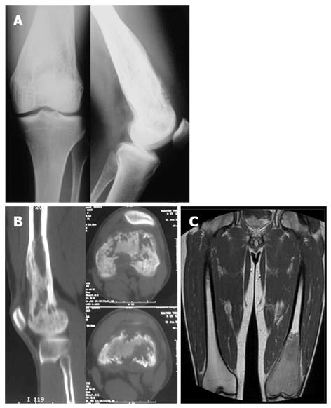
Figure 1 Imaging of the lesion at the patient admission.
A: Lytic lesion of the distal femur is seen on radiographs; B: Computed tomography scan of distal femur showing the erosion of the bone with focal cortical destruction; C: T1-weighted magnetic resonance imaging demonstrating the extent of the lesion with areas of cortical destruction and periostic reaction.
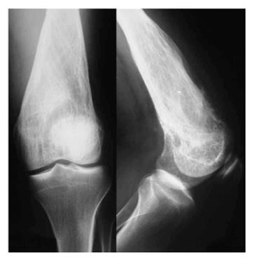
Figure 2 Radiograph at 1 mo follow up.
More obvious lysis of the distal femur comparatively to the initial image. Thick and coarse trabeculation is obviously apparent. Cortical breech is clearly found especially to the anterior cortex.
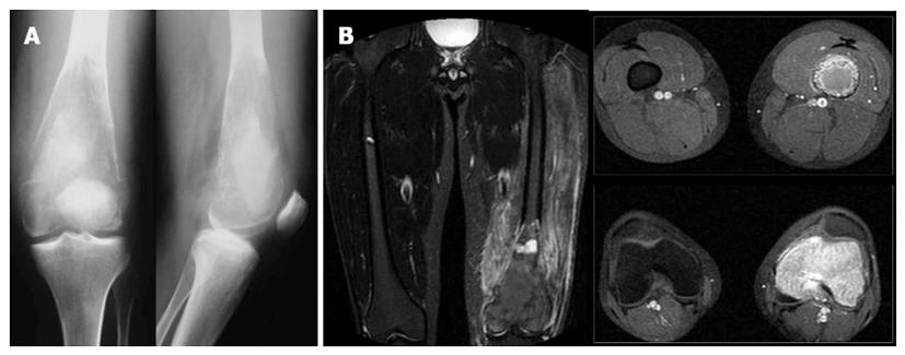
Figure 3 Imaging at three months after the first admission.
A: Elimination of the bone trabeculation is accompanied by a compressive fracture of the metaphysic; B: Magnetic resonance imaging shows no signs of wider extension of the lesion. STIR-weight images reveal an increased oedema of the surrounding muscles.
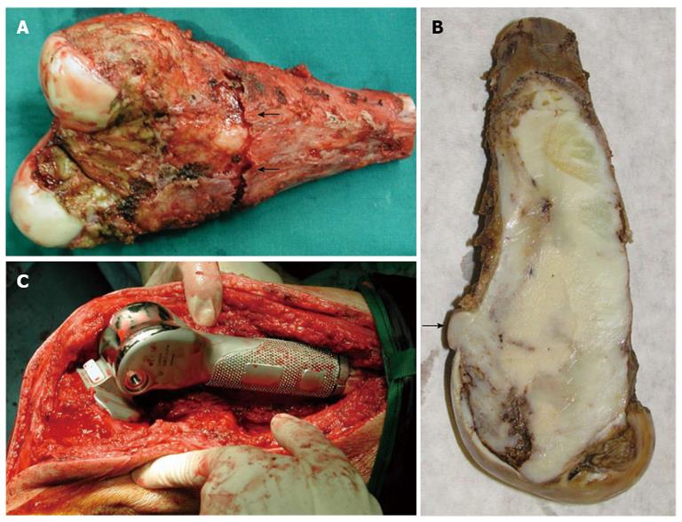
Figure 4 Open biopsy was performed obtaining bone specimens from the outer lateral condyle with 3 cm × 2 cm × 1 cm total dimensions.
A: Excised specimen of the distal 15.5 cm of the femur. Compressive fracture of the posterior cortex (arrows); B: The tumor arose within the medullary cavity and extended from the cartilage surface to 3 cm distance from the excision line. The mass clearly breaches the cortex (arrow); C: Hinged custom made total knee arthroplasty (Howmedica Modular Resection System, Stryker Howmedica Osteonics, Inc).
Figure 5 Microscopic features throughout the tumor.
A: Areas with characteristic irregularly shaped bony spicules, surrounded by hypocellular spindle cell stroma, resembling fibrous dysplasia (HE × 200); B: Focal osteoid production (arrow) within the moderately cellular fibroblastic stroma (HE × 400); C: Occasional mitotic figures were identified (arrow), (HE × 400).
- Citation: Vasiliadis HS, Arnaoutoglou C, Plakoutsis S, Doukas M, Batistatou A, Xenakis TA. Low-grade central osteosarcoma of distal femur, resembling fibrous dysplasia. World J Orthop 2013; 4(4): 327-332
- URL: https://www.wjgnet.com/2218-5836/full/v4/i4/327.htm
- DOI: https://dx.doi.org/10.5312/wjo.v4.i4.327


