Copyright
©2013 Baishideng Publishing Group Co.
World J Orthop. Oct 18, 2013; 4(4): 327-332
Published online Oct 18, 2013. doi: 10.5312/wjo.v4.i4.327
Published online Oct 18, 2013. doi: 10.5312/wjo.v4.i4.327
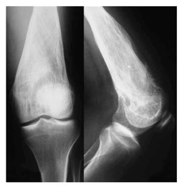
Figure 2 Radiograph at 1 mo follow up.
More obvious lysis of the distal femur comparatively to the initial image. Thick and coarse trabeculation is obviously apparent. Cortical breech is clearly found especially to the anterior cortex.
- Citation: Vasiliadis HS, Arnaoutoglou C, Plakoutsis S, Doukas M, Batistatou A, Xenakis TA. Low-grade central osteosarcoma of distal femur, resembling fibrous dysplasia. World J Orthop 2013; 4(4): 327-332
- URL: https://www.wjgnet.com/2218-5836/full/v4/i4/327.htm
- DOI: https://dx.doi.org/10.5312/wjo.v4.i4.327


