Copyright
©The Author(s) 2022.
World J Orthop. Aug 18, 2022; 13(8): 733-743
Published online Aug 18, 2022. doi: 10.5312/wjo.v13.i8.733
Published online Aug 18, 2022. doi: 10.5312/wjo.v13.i8.733
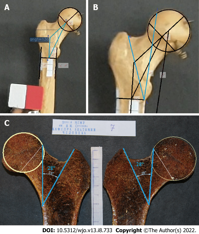
Figure 1 In this study, a method for assessing the morphometric parameters of the proximal femur based on the projection values and angle of rotation around the anatomical axis relative to the FSP was developed.
A: Bone No. 9. Triangle ABC comprised the NSA (angle CAB); AC, neck axis; and AB, diaphyseal axis segment (connecting the diameter of the diaphysis to the neck axis); B: Bone No. 9. Angle KLJ was a part of Ward’s triangle, designating the angle of the minimal resistance zone (AMRZ); C: Bone No. 7. The angle, indicating the borders of the AMRZ, was plotted on photos of gross bone sections.
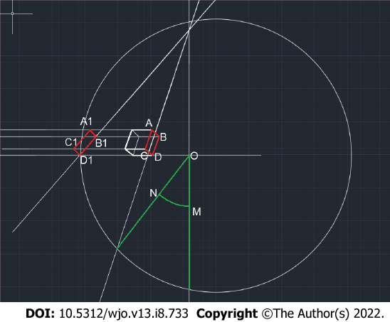
Figure 2 Scheme of the constructions used to determine the angle of rotation of the bone (NOM).
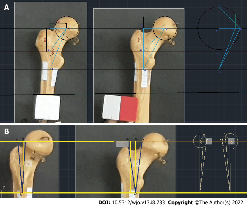
Figure 3 The angle of bone rotation obtained by turning the cube corresponded to the angle measured with the second technique, which uses the previously described feature of the large spit.
A: Bone No. 9. Photo of the NSA and calculation of its true value based on the projection value and the rotation angle; B: Estimation of the true AMRZ based on its projection value and the rotation angle.
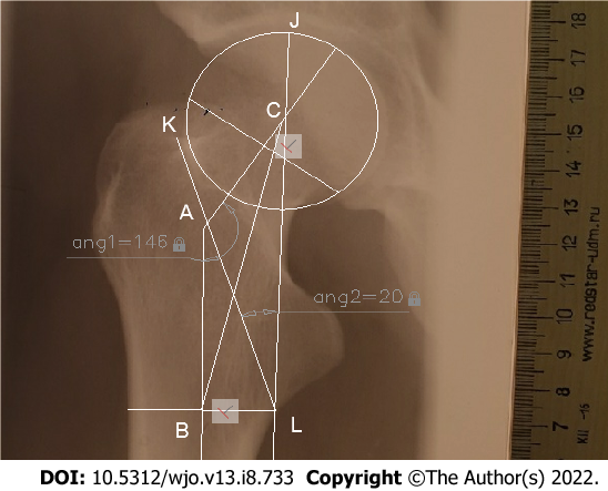
Figure 4 X-ray image from the Department of Human Anatomy showing triangle ABC and the AMRZ.
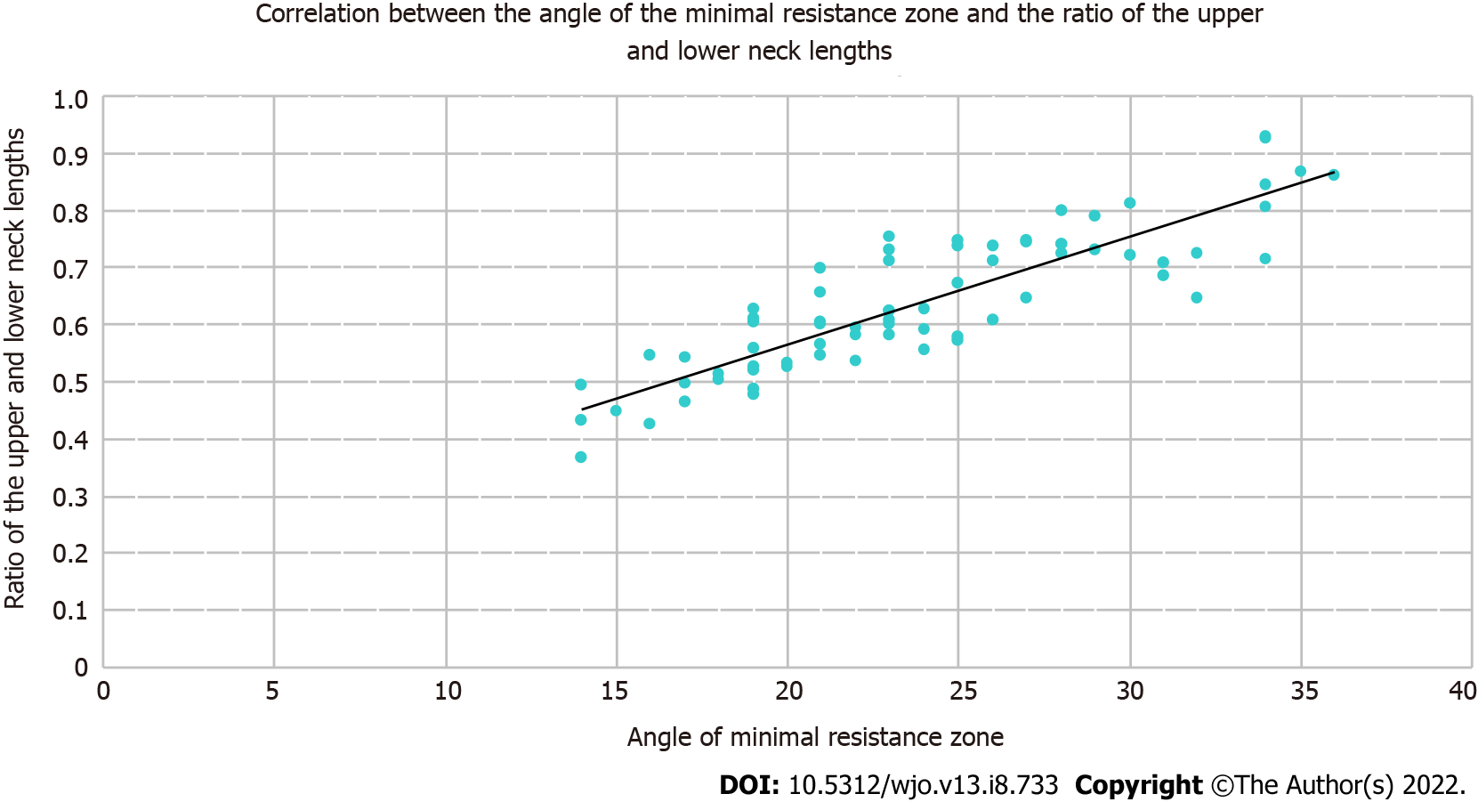
Figure 5 Scatter plot showing the correlation between these parameters and the corresponding regression line.
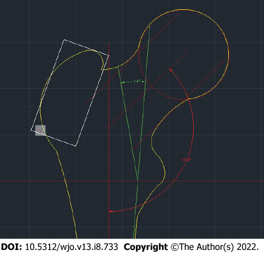
Figure 6 A two-dimensional model of the proximal femoral epiphysis on the frontal standard projection that provides a minimal risk of femoral neck base fracture after a simple mechanical fall (falling from a patient’s own height).
- Citation: Shitova AD, Kovaleva ON, Olsufieva AV, Gadzhimuradova IA, Zubkov DD, Kniazev MO, Zharikova TS, Zharikov YO. Risk modeling of femoral neck fracture based on geometric parameters of the proximal epiphysis. World J Orthop 2022; 13(8): 733-743
- URL: https://www.wjgnet.com/2218-5836/full/v13/i8/733.htm
- DOI: https://dx.doi.org/10.5312/wjo.v13.i8.733


