Copyright
©The Author(s) 2021.
World J Orthop. Sep 18, 2021; 12(9): 629-639
Published online Sep 18, 2021. doi: 10.5312/wjo.v12.i9.629
Published online Sep 18, 2021. doi: 10.5312/wjo.v12.i9.629
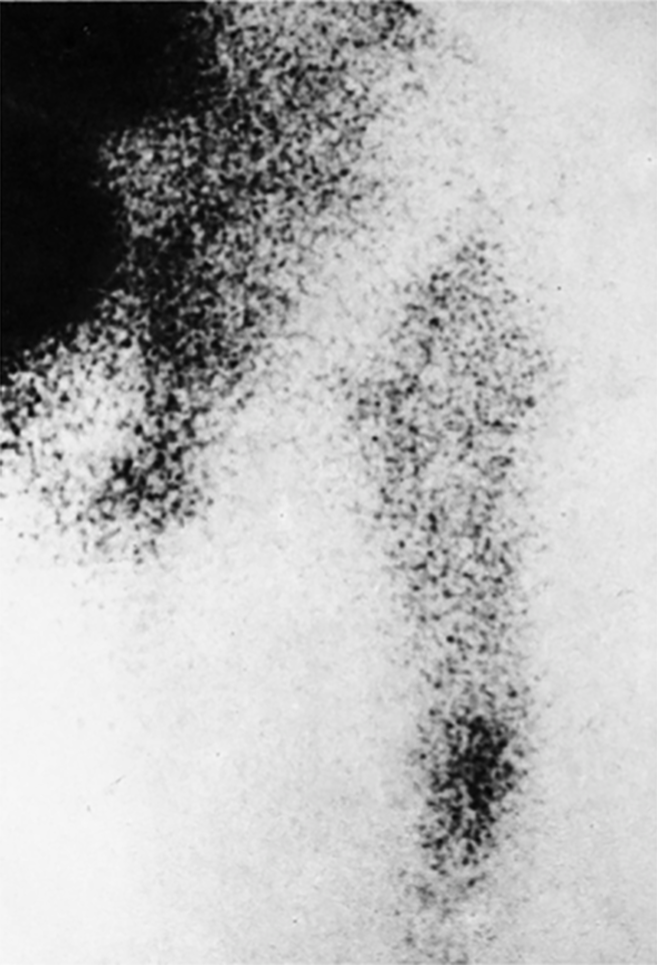
Figure 1 Focally increased activity at the tip of a migrating femoral component at 99mTc-MDP scan.
From Mjöberg et al[16] with permission.
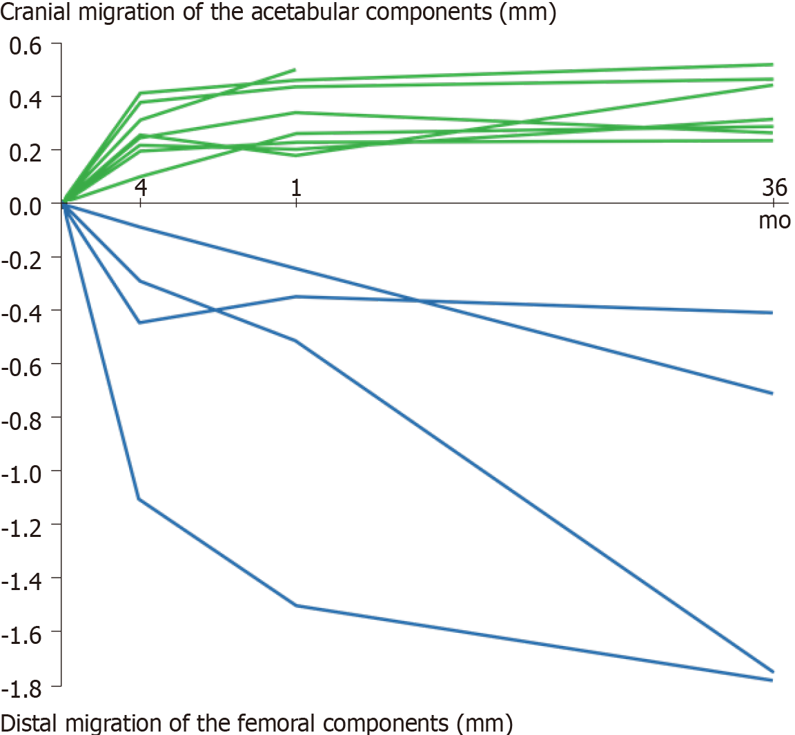
Figure 2 Prosthetic migration along the longitudinal axis.
Migration of the migrating eight acetabular (green) and four femoral components (blue) in the series followed by radiostereometric analysis for 3 years [eight acetabular and ten femoral components did not pass the limit (0.2 mm) for significant migration]. From Mjöberg et al[18] with permission.
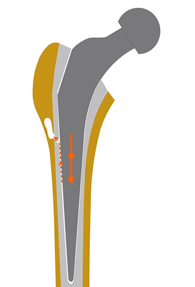
Figure 3 Graph shows how prosthetic micromovements cause a focal osteolysis.
The prosthetic micromovements (orange arrows) pump joint fluid under high pressure (orange dashed arrows) from the gap between the stem and the cement through a defect in the cement mantle. The pressure waves may devitalize the adjacent bone tissue, which is resorbed, thus causing a focal osteolysis. From Mjöberg[53] with permission.
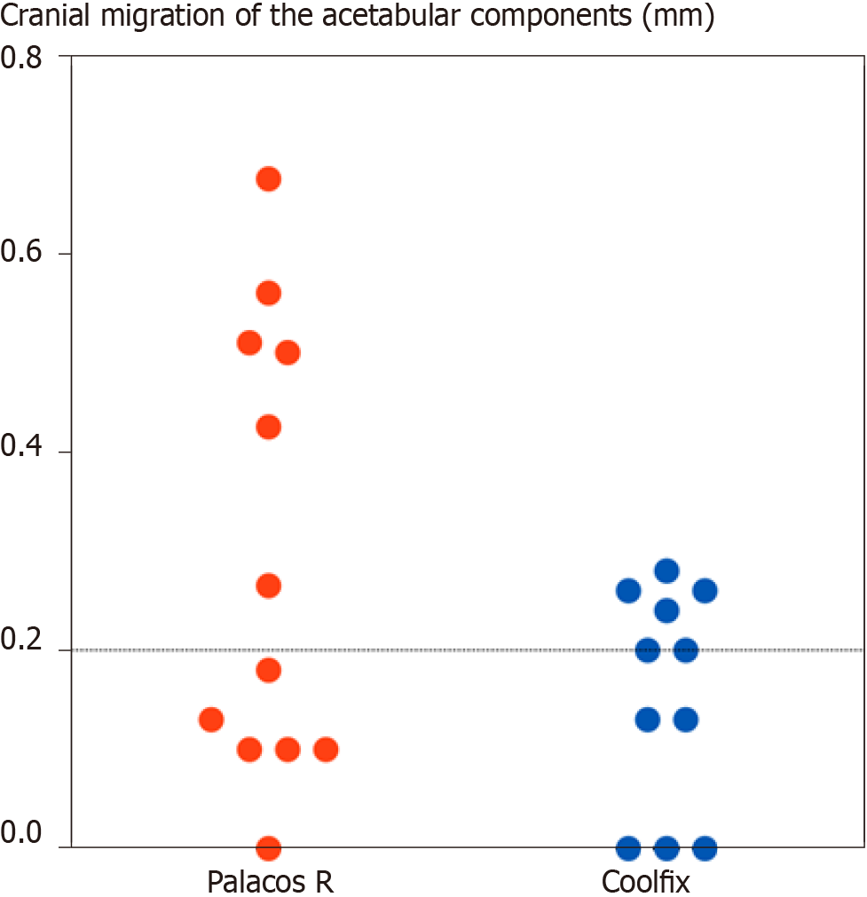
Figure 4 Cranial migration of the acetabular components after 1 year.
By then, six of the 12 with Palacos R and four of the 11 with Coolfix had passed the limit (0.2 mm) for significant migration. However, five acetabular components with Palacos R had migrated 0.4–0.7 mm, while all 11 with Coolfix had migrated less than 0.3 mm (P < 0.04, Student’s t-test, one-tailed).
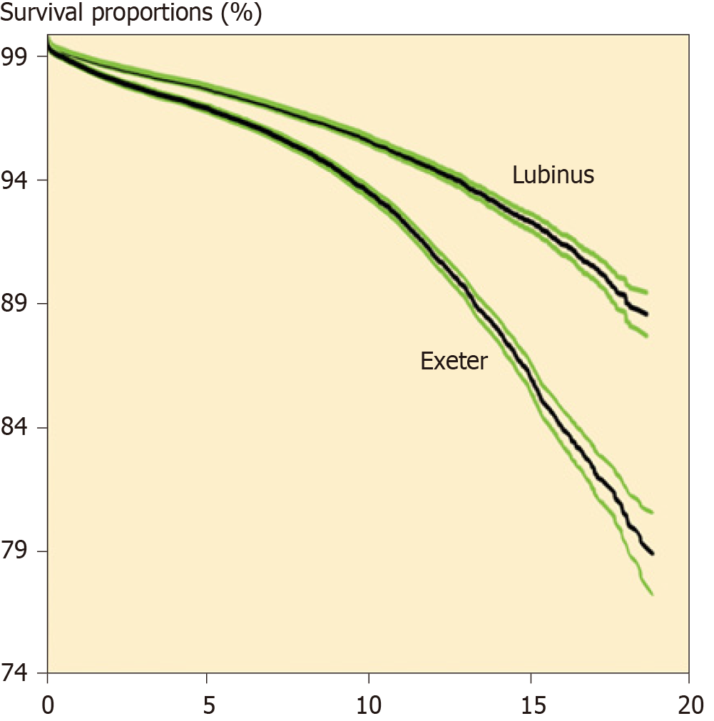
Figure 5 Kaplan-Meier implant survival of cemented total hip devices.
Calculated from the Nordic Arthroplasty Register Association database. Green lines are upper and lower 95% confidence limits. Compiled from Junnila et al[64] with permission.
- Citation: Mjöberg B. Hip prosthetic loosening: A very personal review. World J Orthop 2021; 12(9): 629-639
- URL: https://www.wjgnet.com/2218-5836/full/v12/i9/629.htm
- DOI: https://dx.doi.org/10.5312/wjo.v12.i9.629


