Copyright
©The Author(s) 2021.
World J Orthop. Apr 18, 2021; 12(4): 214-222
Published online Apr 18, 2021. doi: 10.5312/wjo.v12.i4.214
Published online Apr 18, 2021. doi: 10.5312/wjo.v12.i4.214
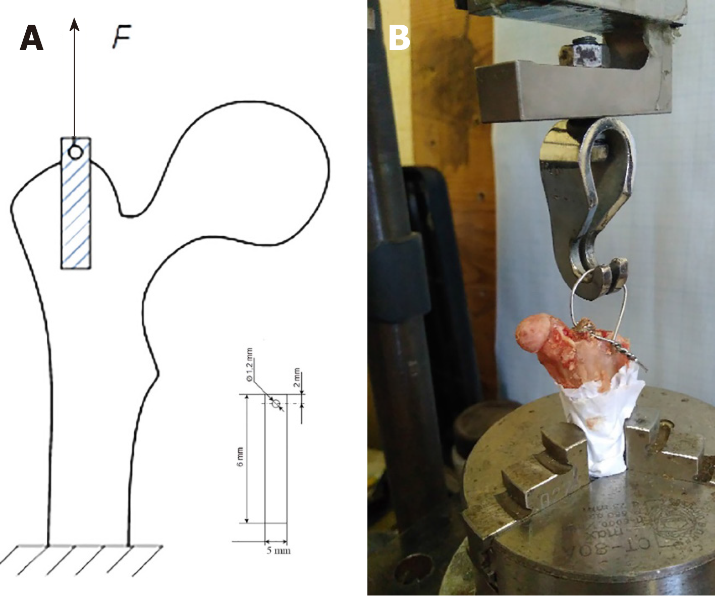
Figure 1 Scheme of biomechanical testing and biomechanical testing.
A: The geometric dimension of the implants; B: Rabbit femur with implant on the stand during testing.
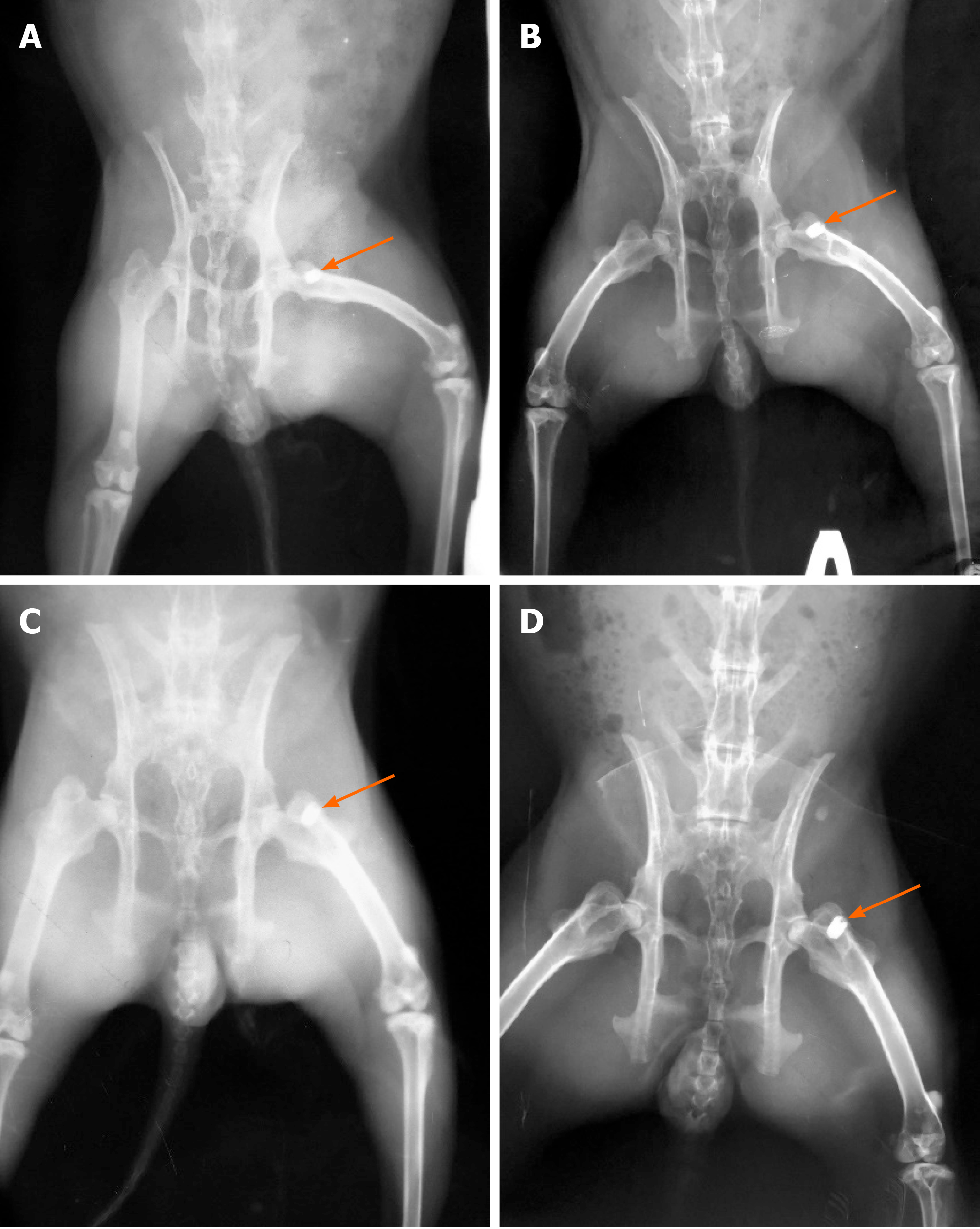
Figure 2 Х-ray images of rabbits after implanted CONCELOC material (arrow) in the proximal femur.
А: Non-ovariectomized (non-OVX) rabbit after surgery; B: Non-OVX rabbit at 8 wk after surgery; C: Ovariectomized (OVX) rabbit after surgery; D: OVX rabbit at 8 wk after surgery.
Figure 3 Evaluation of the cortical thickness index of the proximal femur of rabbit with "X-Rays" software (Kharkiv National University of Radioelectronics, Ukraine) 3 mo after ovariectomy.
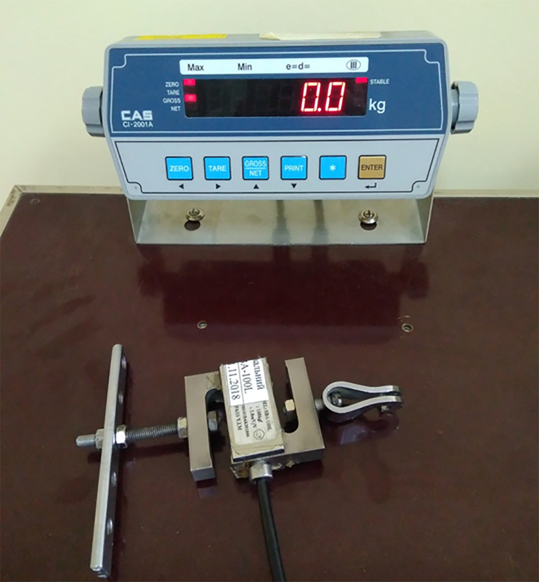
Figure 4 Device for recording breakout force with a strain gauge.
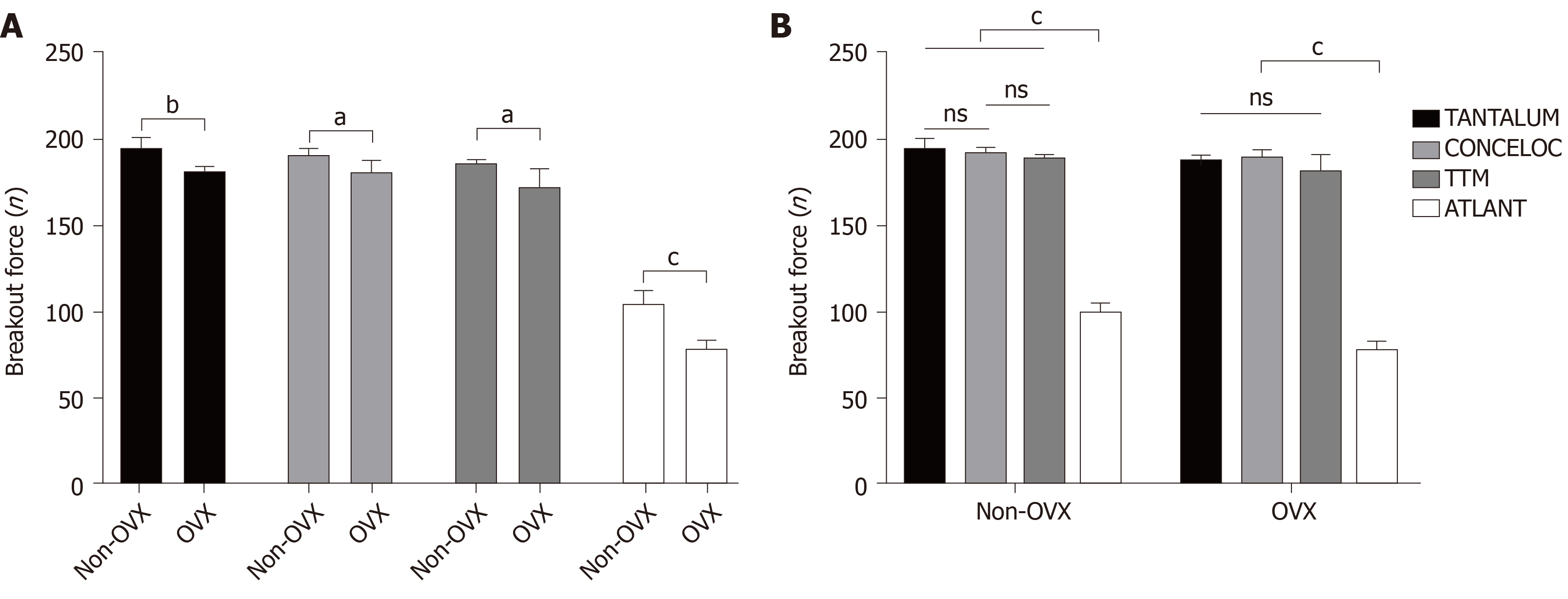
Figure 5 Results of breakout force testing of four types of porous materials in ovariectomized (OVX, n = 20) and healthy rabbits (non-OVX, n = 20) 8 wk after implantation.
А: Unpaired t-test: Analysis of the effect of osteoporosis (OVX group) on the bone-implant strength and osseointegration for the same type of implant; B: ANOVA with post-hoc Duncan test evaluation of the effect of the implant material on bone-implant strength and osseointegration in ovariectomized and non-ovariectomized rabbits. aP < 0.05; bP < 0.01; cP < 0.001. ns: not significant; non-OVX: non-ovariectomized; OVX: ovariectomized.
- Citation: Bondarenko S, Filipenko V, Karpinsky M, Karpinska O, Ivanov G, Maltseva V, Badnaoui AA, Schwarzkopf R. Osseointegration of porous titanium and tantalum implants in ovariectomized rabbits: A biomechanical study. World J Orthop 2021; 12(4): 214-222
- URL: https://www.wjgnet.com/2218-5836/full/v12/i4/214.htm
- DOI: https://dx.doi.org/10.5312/wjo.v12.i4.214


