Copyright
©The Author(s) 2020.
World J Orthop. Aug 18, 2020; 11(8): 345-356
Published online Aug 18, 2020. doi: 10.5312/wjo.v11.i8.345
Published online Aug 18, 2020. doi: 10.5312/wjo.v11.i8.345
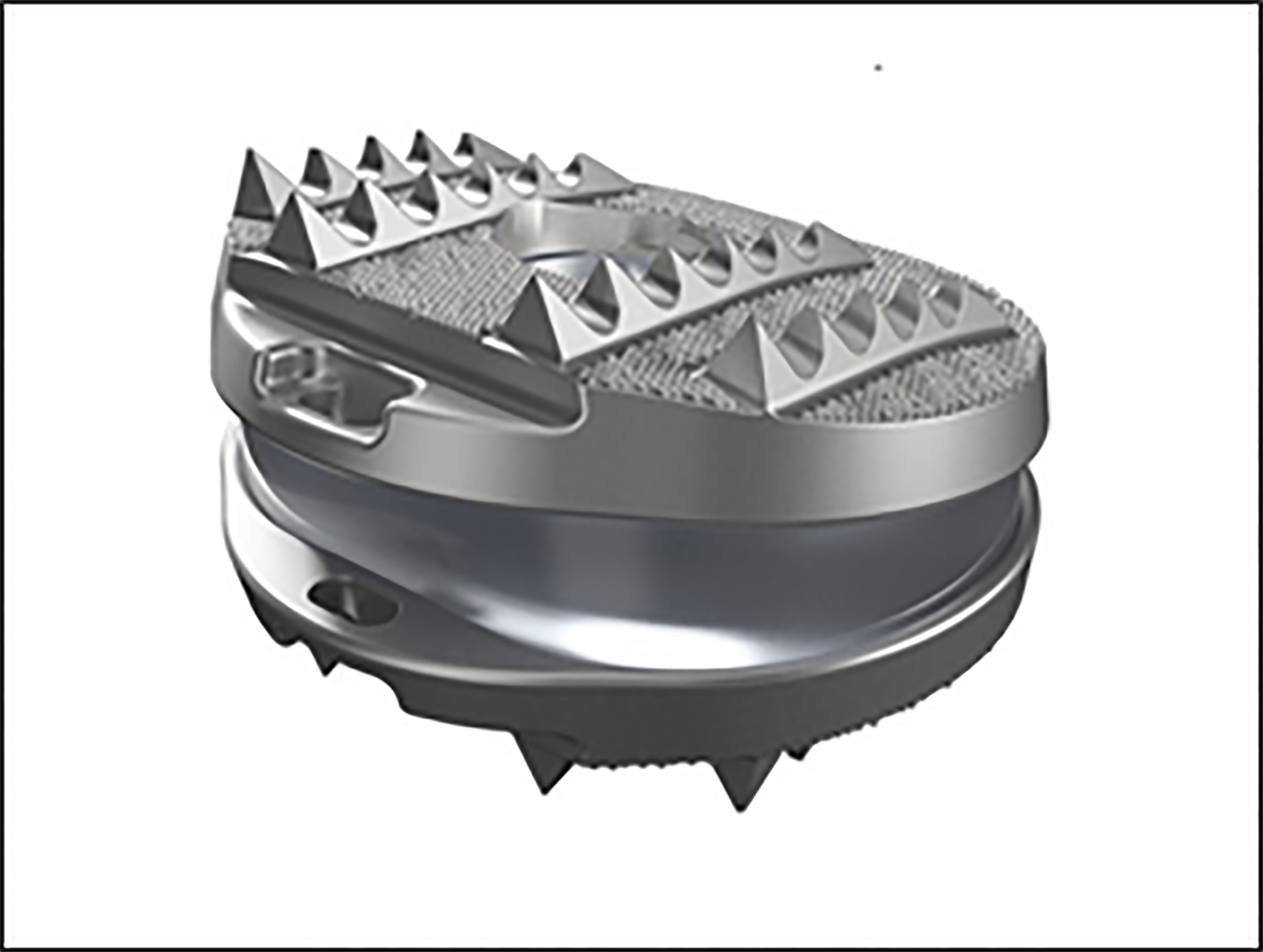
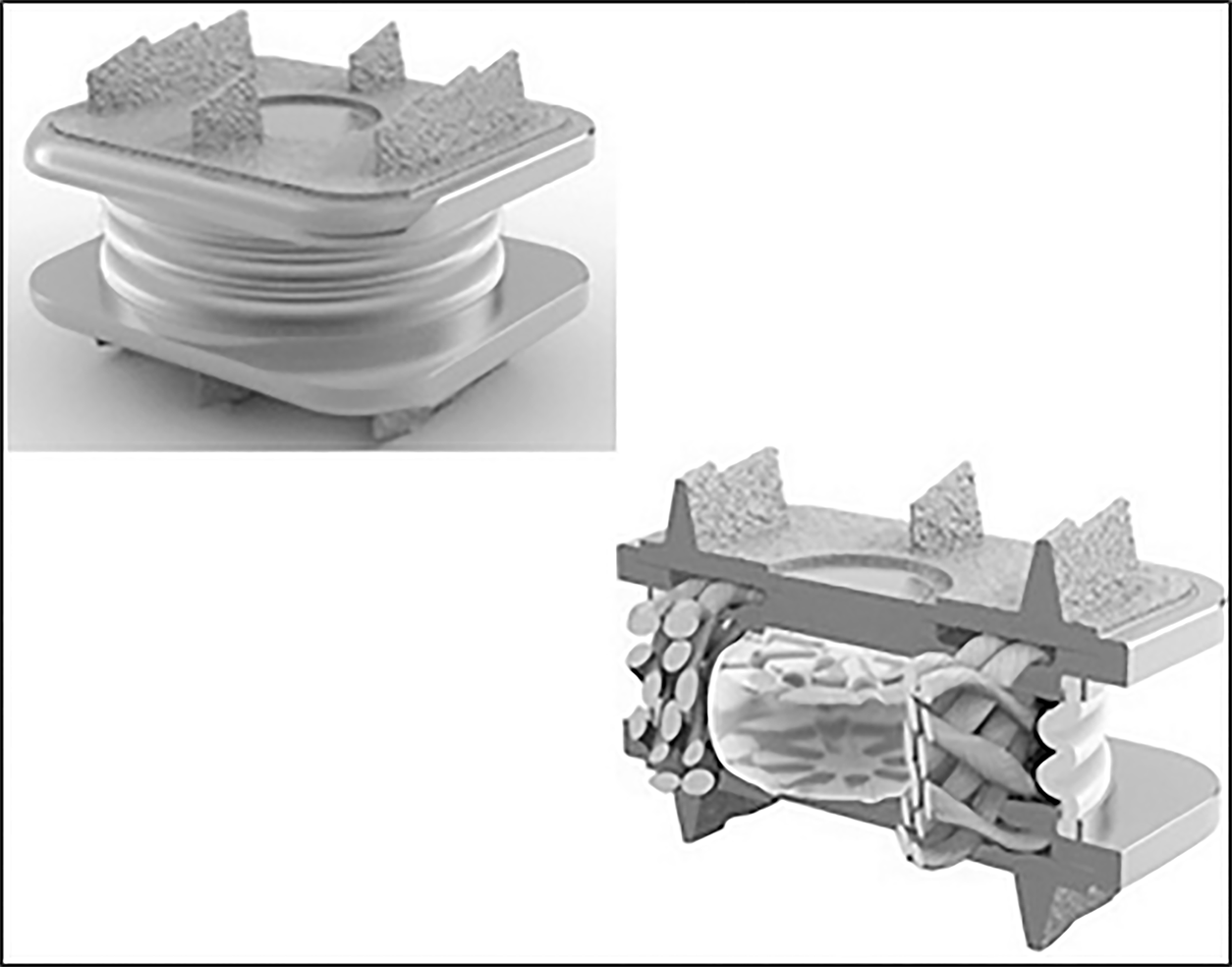
Figure 1 Freedom lumbar disc.
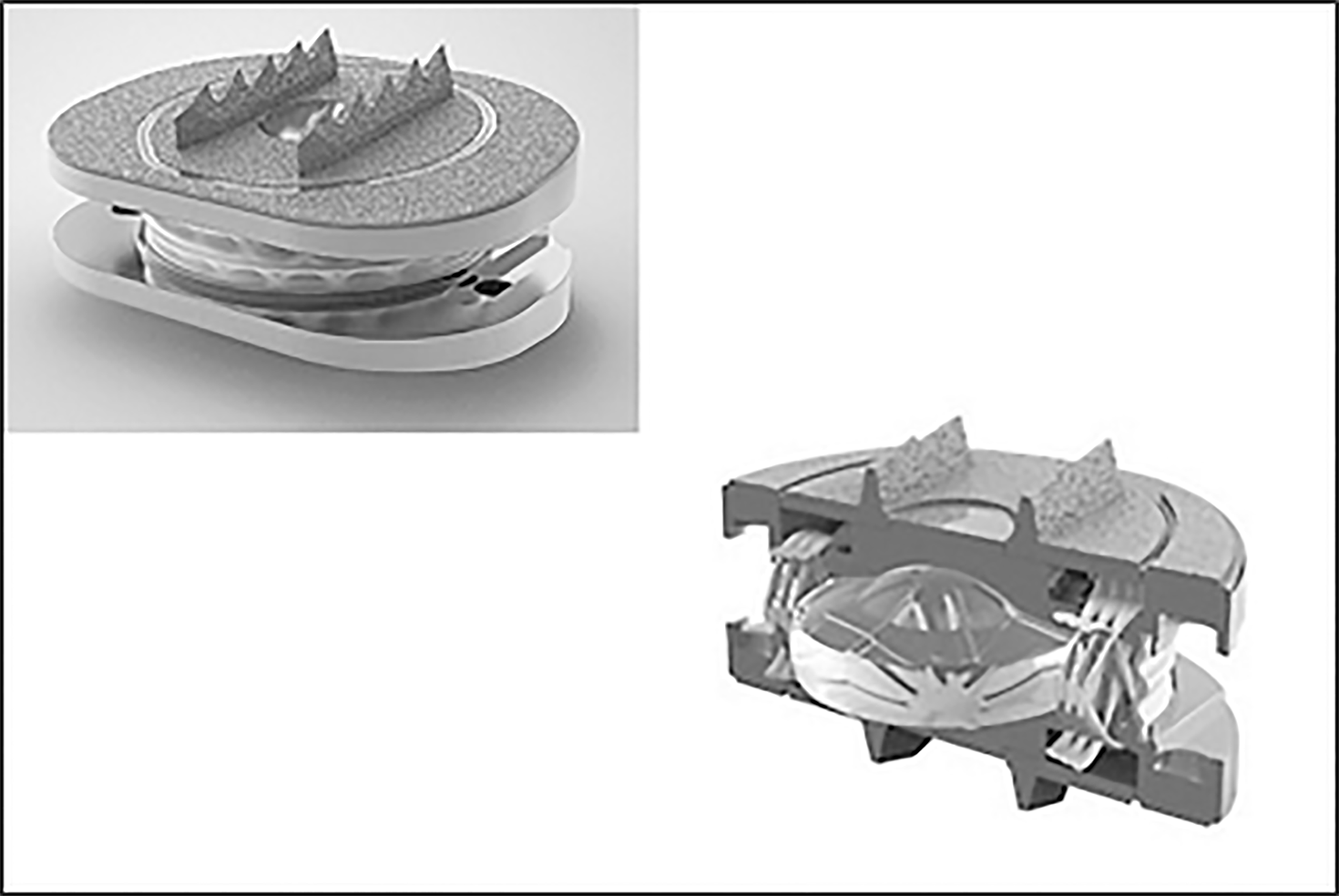
Figure 2 M6-L lumbar disc.
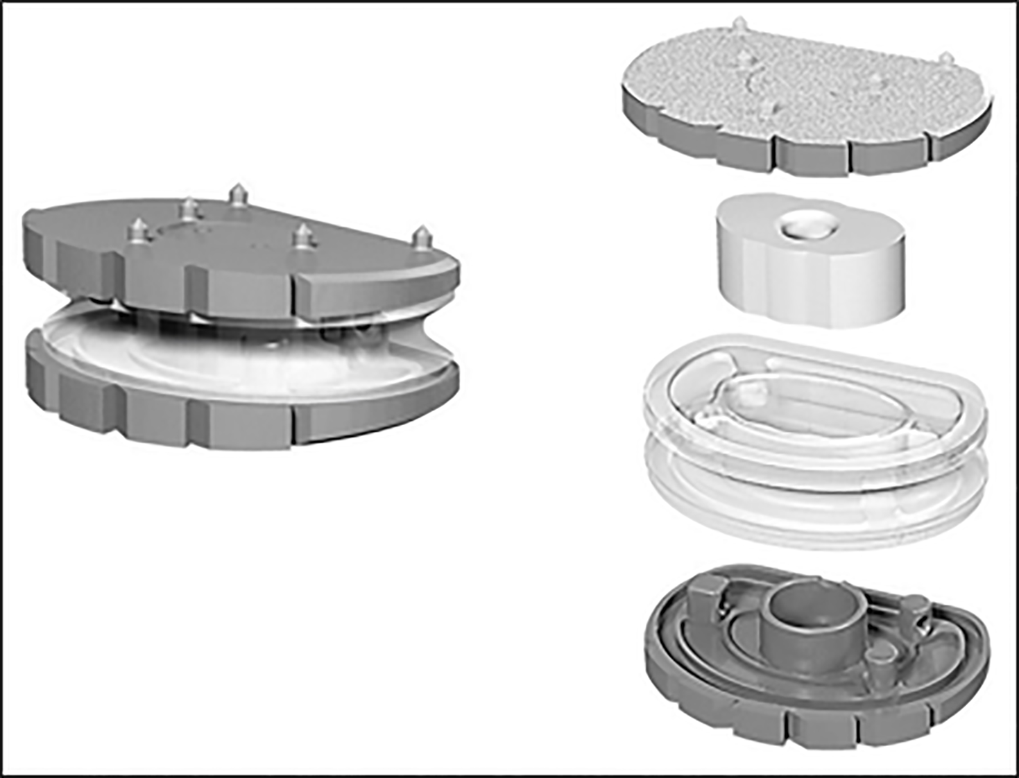
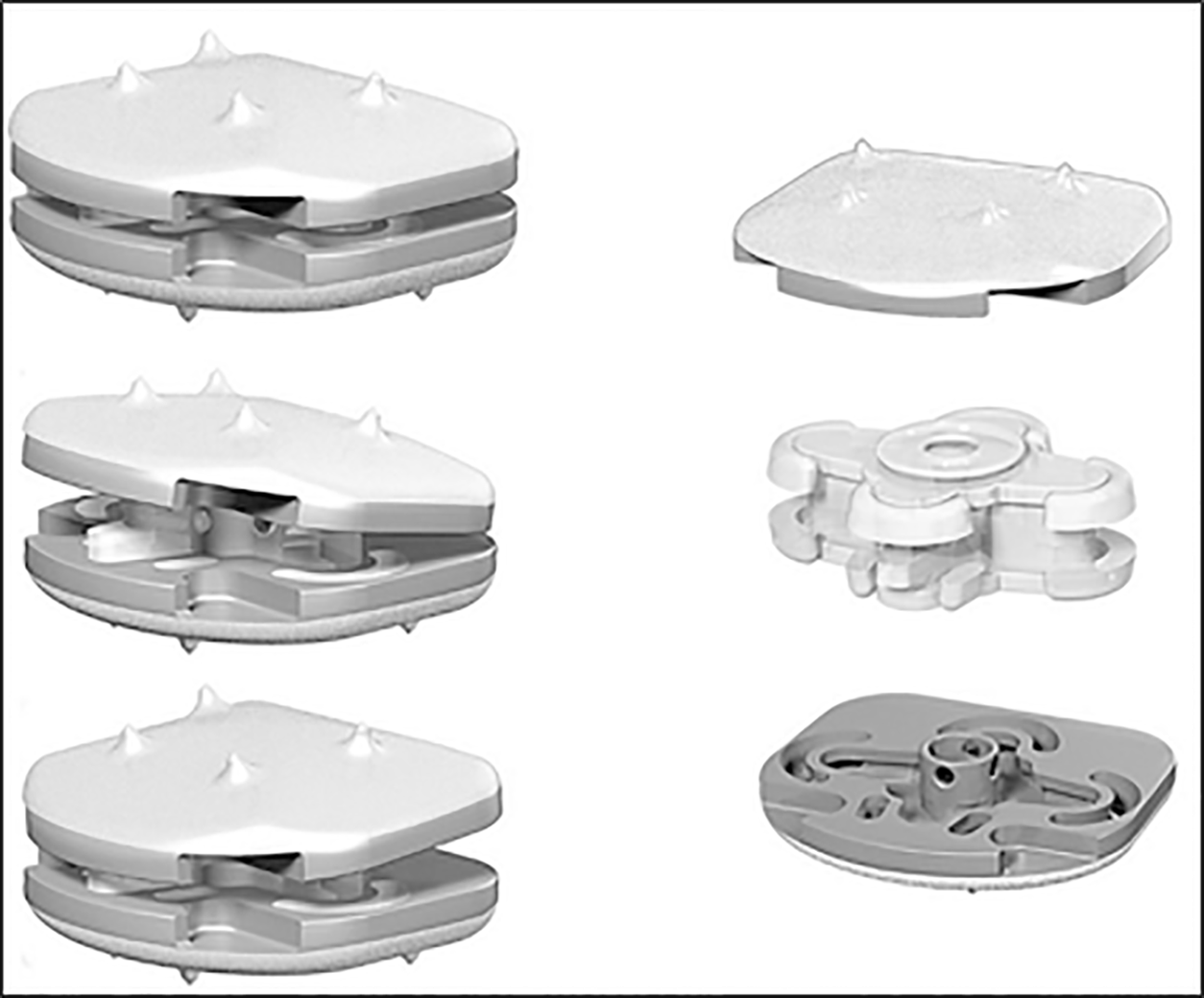
Figure 3 LP ESP lumbar disc.
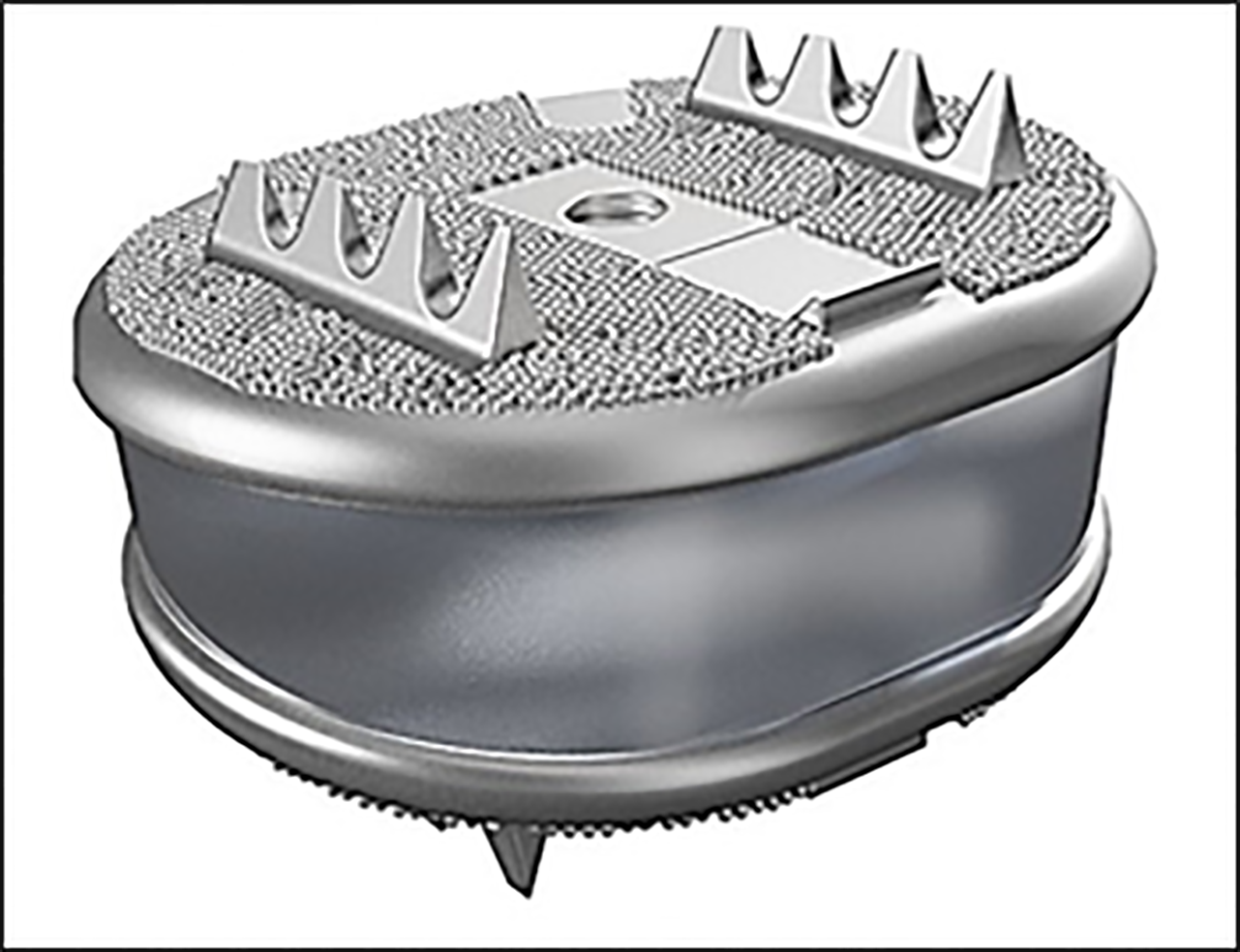
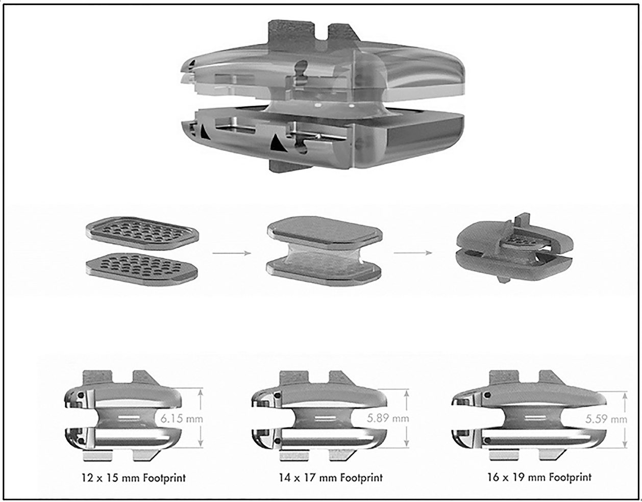
Figure 4 Freedom cervical disc.
Figure 5 M6-C cervical disc.
Figure 6 CP-ESP cervical disc.
Figure 7 Rhine cervical disc.
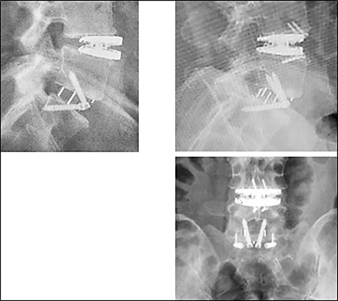
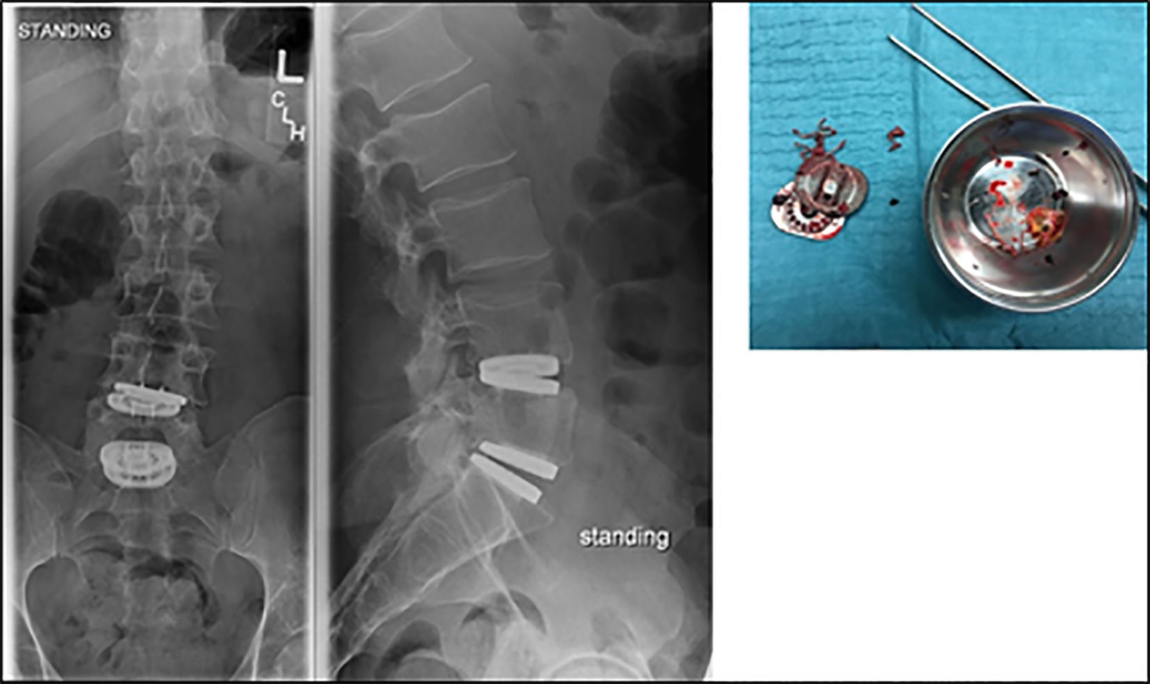
Figure 8 L4L5 LP ESP lumbar disc migration.
Insufficient primary fixation and early anterior migration. Combined L5S1 ALIF. The ESP implant was repositioned by impaction during revision surgery. The surgeon used small screws as an anterior stop to stabilize the implant until effective bone growth and secondary fixation.
Figure 9 M6-L lumbar disc failure.
Rupture of the ultrahigh-molecular weight polyethylene fibers construct and the peripheral polymer sheath. Nucleus expulsion and direct endplates contact with local metallosis.
- Citation: Lazennec JY. Lumbar and cervical viscoelastic disc replacement: Concepts and current experience. World J Orthop 2020; 11(8): 345-356
- URL: https://www.wjgnet.com/2218-5836/full/v11/i8/345.htm
- DOI: https://dx.doi.org/10.5312/wjo.v11.i8.345


