Copyright
©The Author(s) 2020.
World J Orthop. Feb 18, 2020; 11(2): 116-122
Published online Feb 18, 2020. doi: 10.5312/wjo.v11.i2.116
Published online Feb 18, 2020. doi: 10.5312/wjo.v11.i2.116
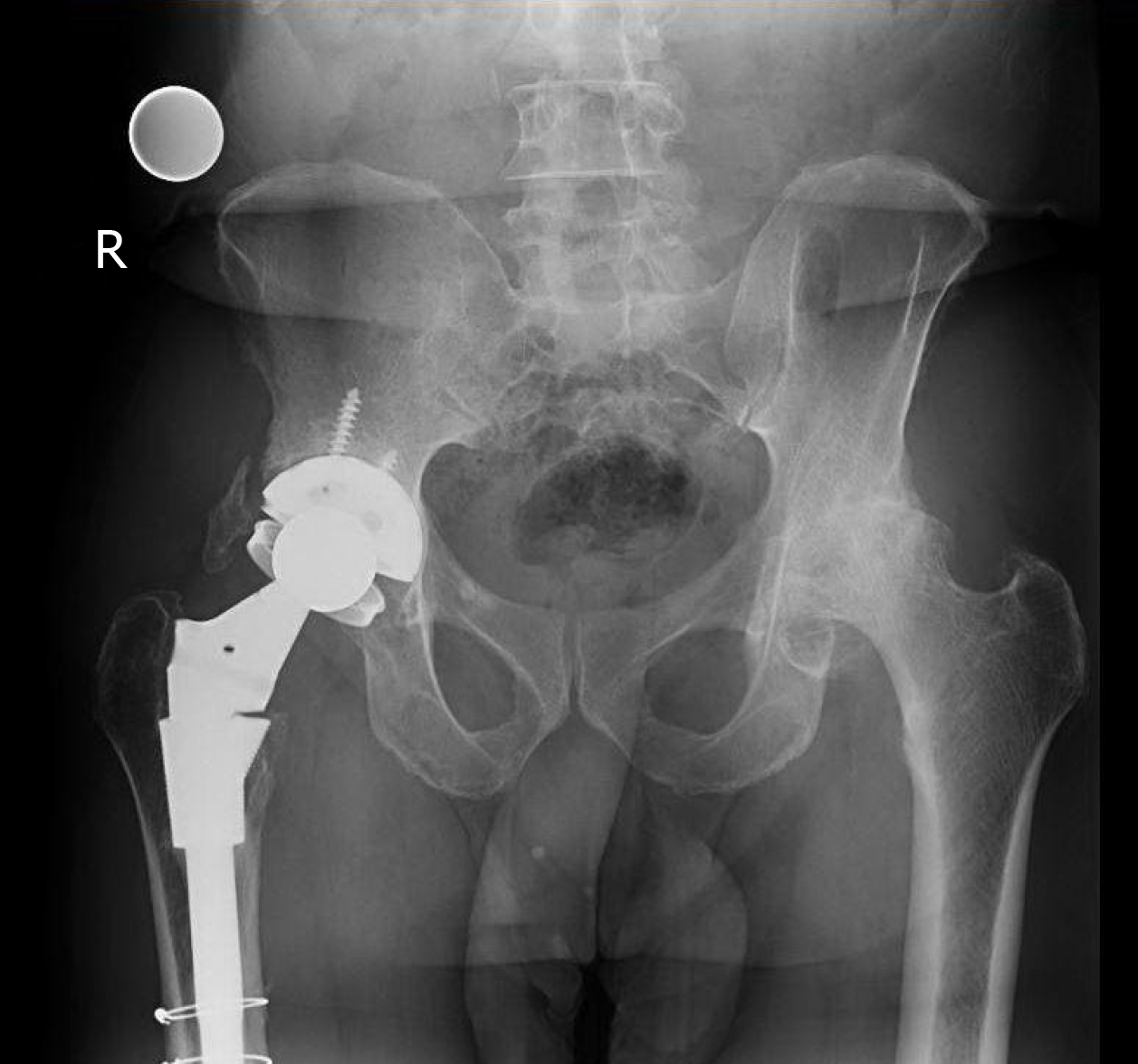
Figure 1 Pre-operative anterior-posterior pelvic radiograph showing degenerative changes of left hip with severe joint space narrowing, subchondral sclerosis, and cyst formation.
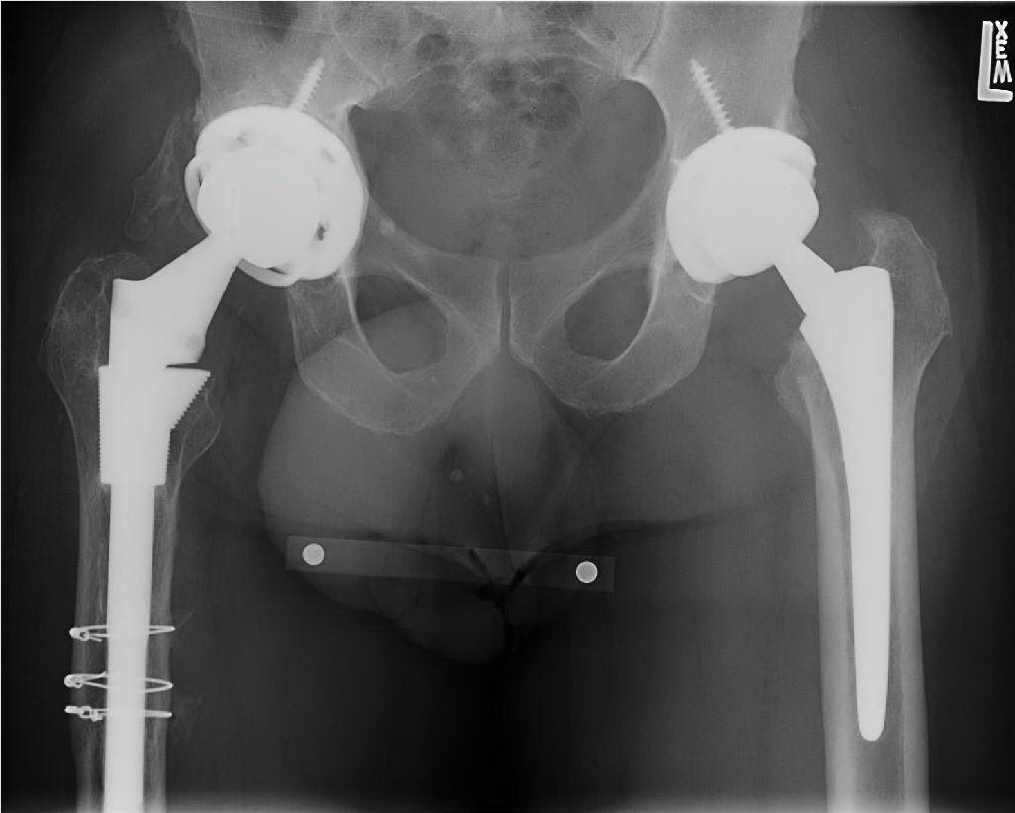
Figure 2 Initial post-operative anterior-posterior pelvic radiograph showing the components of the prosthesis well-aligned and well seated.
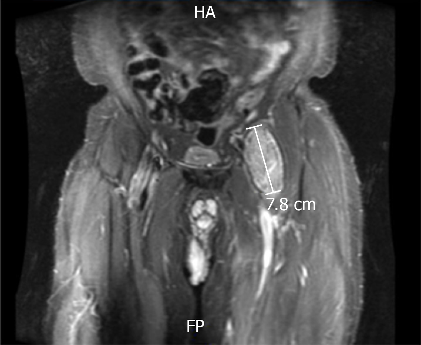
Figure 3 Pre-revision T2-weighted coronal magnetic resonance image showing a well-defined 7.
8 cm complex lesion anterior to the left hip prosthesis.
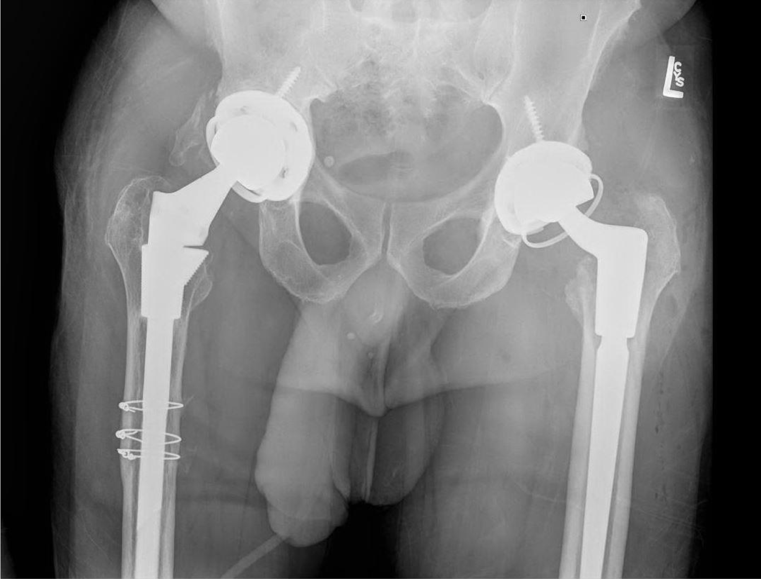
Figure 4 Post-revision anterior-posterior hip radiograph showing an aligned and well-fixed left hip prosthesis.
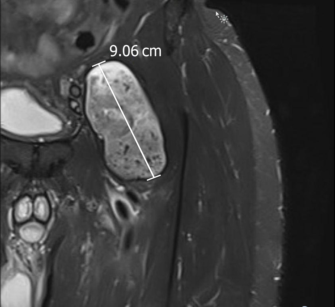
Figure 5 Post-revision short-T1 inversion recovery coronal magnetic resonance image demonstrating pseudotumor recurrence.
- Citation: Desai BR, Sumarriva GE, Chimento GF. Pseudotumor recurrence in a post-revision total hip arthroplasty with stem neck modularity: A case report. World J Orthop 2020; 11(2): 116-122
- URL: https://www.wjgnet.com/2218-5836/full/v11/i2/116.htm
- DOI: https://dx.doi.org/10.5312/wjo.v11.i2.116


