Copyright
©The Author(s) 2020.
World J Orthop. Dec 18, 2020; 11(12): 615-626
Published online Dec 18, 2020. doi: 10.5312/wjo.v11.i12.615
Published online Dec 18, 2020. doi: 10.5312/wjo.v11.i12.615
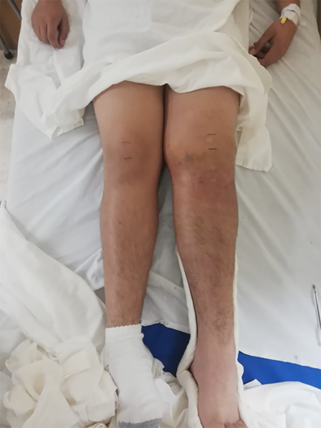
Figure 1 Preoperative clinical photograph of the patient's lower extremities, where the swelling in the affected knee (left) can be observed, as well as asymmetry in the height of the patella.
The upper and lower margins of the patella were marked with a pen.
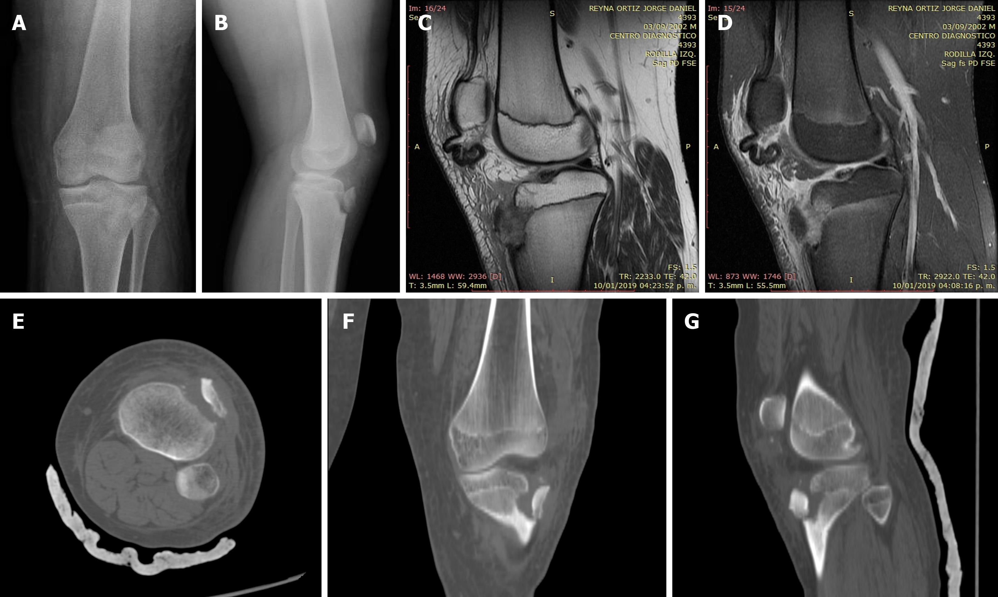
Figure 2 Patient imaging studies.
A and B: Simple radiographs in the two standard projections (anteroposterior and lateral), where the avulsion fracture of the TT is observed without apparent involvement of the articular surface; C and D: Sagittal sections of magnetic resonance images with T1 and T2 sequences, where the distal rupture and proximal retraction of the patellar tendon can be observed, maintaining its integrity; E-G: Computed tomography images taken in the axial, coronal and sagittal views of the bone window, where the displacement of the bone fragment, the eversion and 180º degrees of rotation of the same segment can be observed; the anterior cortex rotated posteriorly, confirming the integrity of the articular surface.
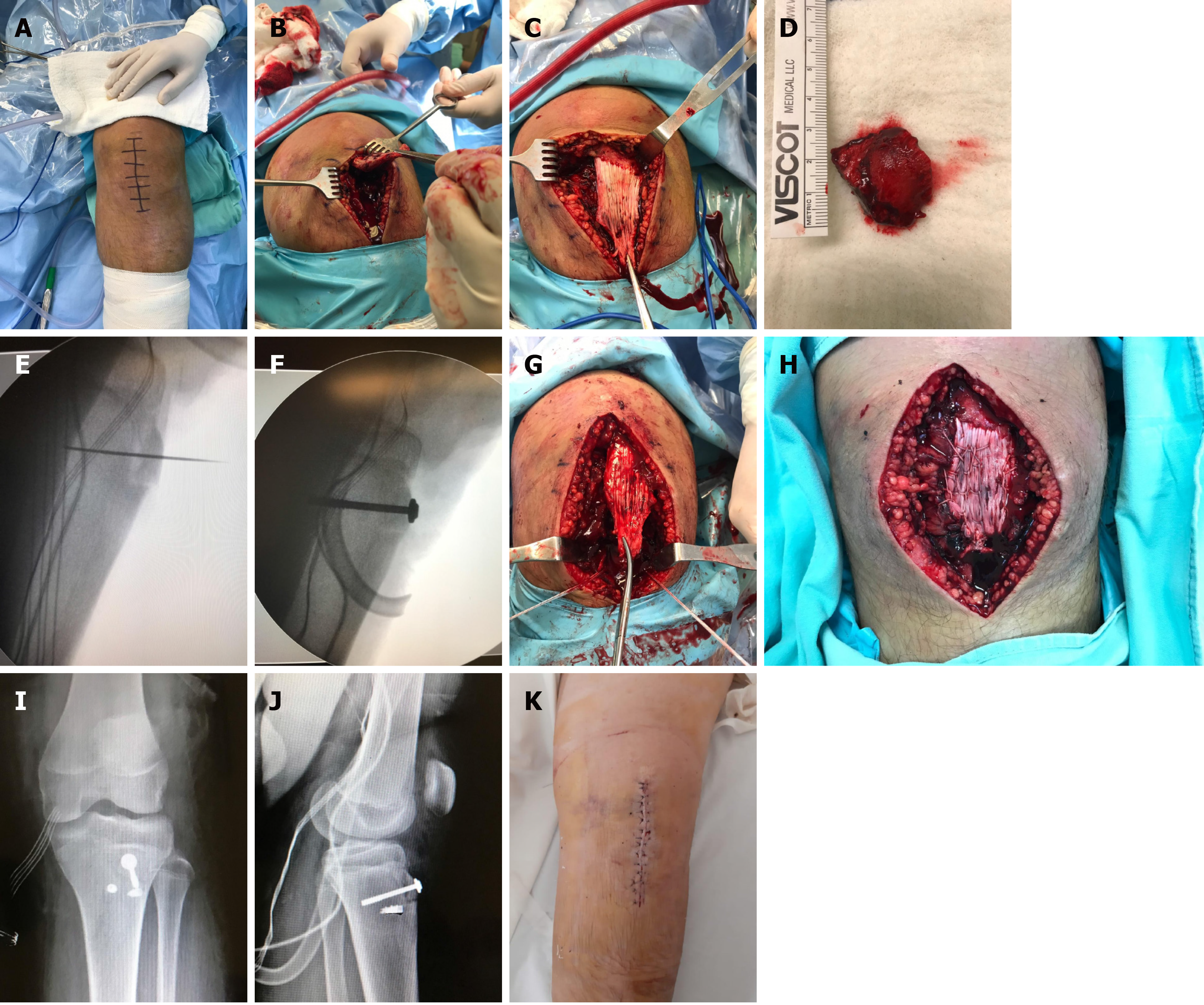
Figure 3 Surgical procedure performed.
A: The incision made; B: Identification of the proximally retracted patellar tendon; C: Image showing its integrity; D: The only large avulsed bone fragment; E: Reduction and temporal fixation of the bone fragment with K-wire; F: Definitive osteosynthesis with the use of a cannulated screw with a partial thread of 6.5 mm in diameter and the use of a washer; G: The placement of two steel anchors on each side of the newly fixed tibial tuberosity; H: Final appearance of the reinserted tendon; I and J: Immediate postoperative radiographs in two projections; K: Final appearance of the knee at 24 h postoperatively.
- Citation: Morales-Avalos R, Martínez-Manautou LE, de la Garza-Castro S, Pozos-Garza AJ, Villarreal-Villareal GA, Peña-Martínez VM, Vílchez-Cavazos F. Tibial tuberosity avulsion-fracture associated with complete distal rupture of the patellar tendon: A case report and review of literature. World J Orthop 2020; 11(12): 615-626
- URL: https://www.wjgnet.com/2218-5836/full/v11/i12/615.htm
- DOI: https://dx.doi.org/10.5312/wjo.v11.i12.615


