Copyright
©The Author(s) 2020.
World J Orthop. Dec 18, 2020; 11(12): 615-626
Published online Dec 18, 2020. doi: 10.5312/wjo.v11.i12.615
Published online Dec 18, 2020. doi: 10.5312/wjo.v11.i12.615
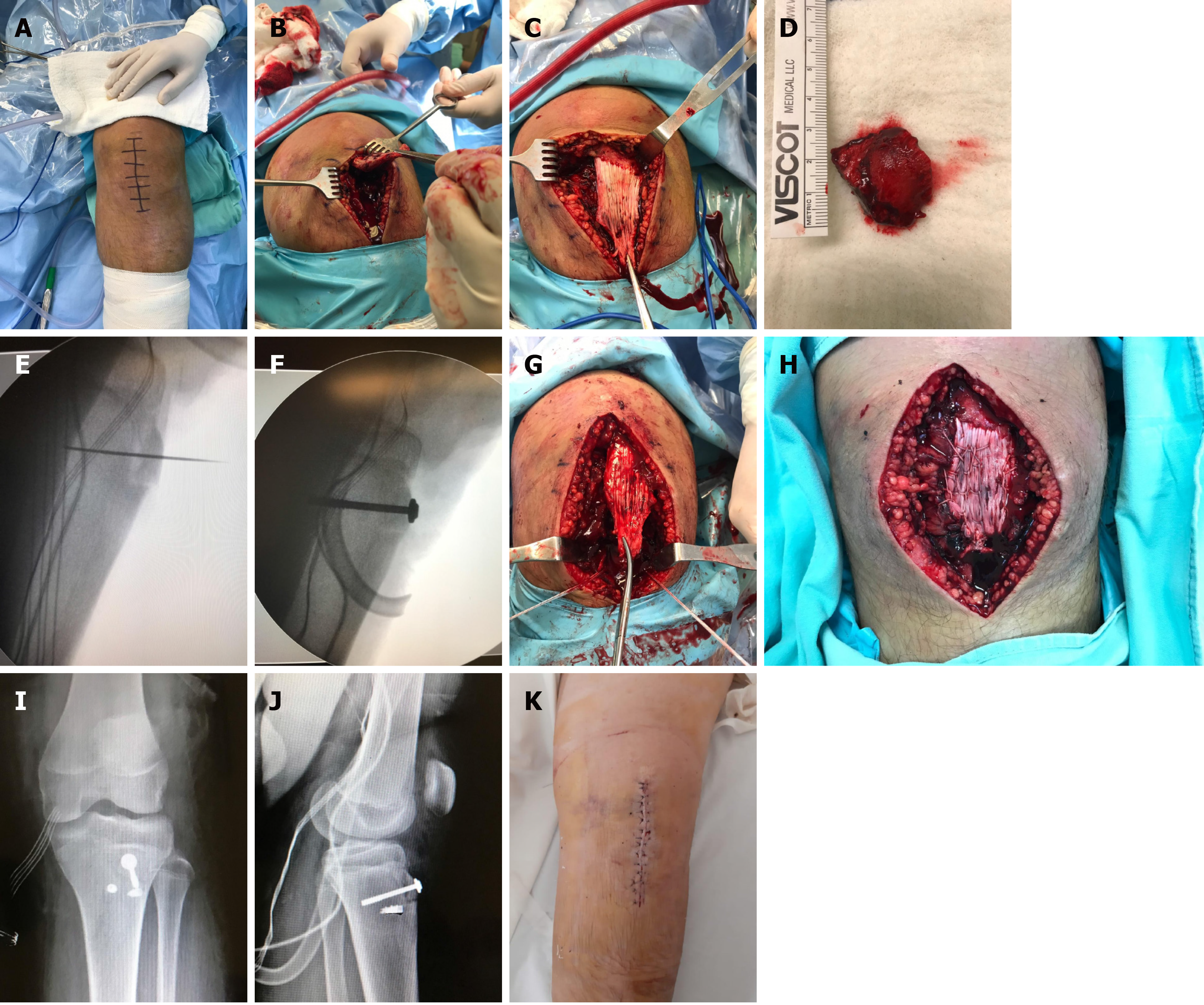
Figure 3 Surgical procedure performed.
A: The incision made; B: Identification of the proximally retracted patellar tendon; C: Image showing its integrity; D: The only large avulsed bone fragment; E: Reduction and temporal fixation of the bone fragment with K-wire; F: Definitive osteosynthesis with the use of a cannulated screw with a partial thread of 6.5 mm in diameter and the use of a washer; G: The placement of two steel anchors on each side of the newly fixed tibial tuberosity; H: Final appearance of the reinserted tendon; I and J: Immediate postoperative radiographs in two projections; K: Final appearance of the knee at 24 h postoperatively.
- Citation: Morales-Avalos R, Martínez-Manautou LE, de la Garza-Castro S, Pozos-Garza AJ, Villarreal-Villareal GA, Peña-Martínez VM, Vílchez-Cavazos F. Tibial tuberosity avulsion-fracture associated with complete distal rupture of the patellar tendon: A case report and review of literature. World J Orthop 2020; 11(12): 615-626
- URL: https://www.wjgnet.com/2218-5836/full/v11/i12/615.htm
- DOI: https://dx.doi.org/10.5312/wjo.v11.i12.615


