Copyright
©The Author(s) 2019.
World J Orthop. Oct 18, 2019; 10(10): 371-377
Published online Oct 18, 2019. doi: 10.5312/wjo.v10.i10.371
Published online Oct 18, 2019. doi: 10.5312/wjo.v10.i10.371
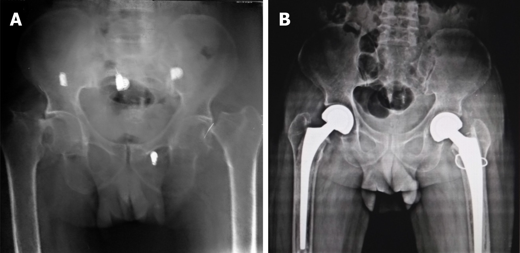
Figure 1 Radiograph.
A: Preoperative radiograph showing bilateral displaced fracture neck of femur with significant osteopenia. B: Postoperative radiograph at three months. There was a fracture of the greater trochanter on the left side which was fixed with stainless steel wire. There is a good union of the greater trochanter.
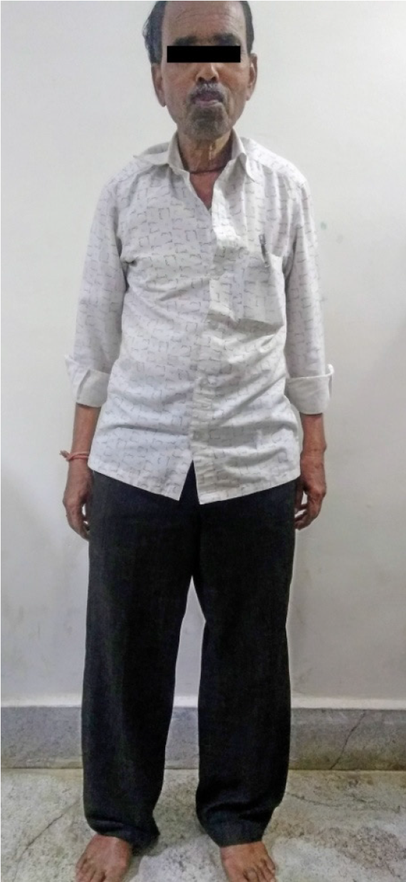
Figure 2 Patient walking unsupported at three months after the surgery.
- Citation: Sadiq M, Kulkarni V, Hussain SA, Ismail M, Nayak M. Low-velocity simultaneous bilateral femoral neck fracture following long-term antiepileptic therapy: A case report. World J Orthop 2019; 10(10): 371-377
- URL: https://www.wjgnet.com/2218-5836/full/v10/i10/371.htm
- DOI: https://dx.doi.org/10.5312/wjo.v10.i10.371


