Copyright
©2010 Baishideng Publishing Group Co.
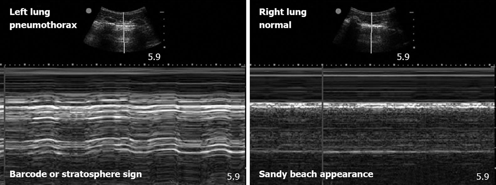
Figure 1 Pneumothorax on bedside ultrasonography.
These two images utilize M-mode echography to gather more information on the pleural interfaces seen as a hyperechoic line in the B-mode images above. The M-mode tracings below represent a single line of sight through the B-mode images. Over time, the gray-scale appearance of M-mode images changes with the movement of the tissue. In normal lungs the pleurae slide across each other and this movement takes on a “granular” or “sandy beach” appearance (image on the right). As a result of the lack of pleural sliding in the presence of pneumothorax, a pattern of horizontal striations that do not demonstrate the granular appearance of movement is present. This M-mode pattern that is characteristic of pneumothorax has been described as a “barcode” or the “stratosphere” sign (image on the left).
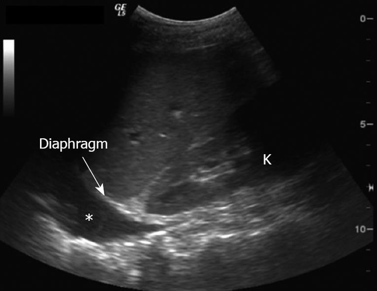
Figure 2 Hemothorax on bedside ultrasonography.
Hemothorax can be seen as hypoechoic area (asterisk) above the liver. Of note, movement of the lung within the pleural fluid can often be appreciated during the progression of the respiratory cycle. The kidney (K) and the diaphragm (arrow) can be easily seen, with the wedge of the liver located above.
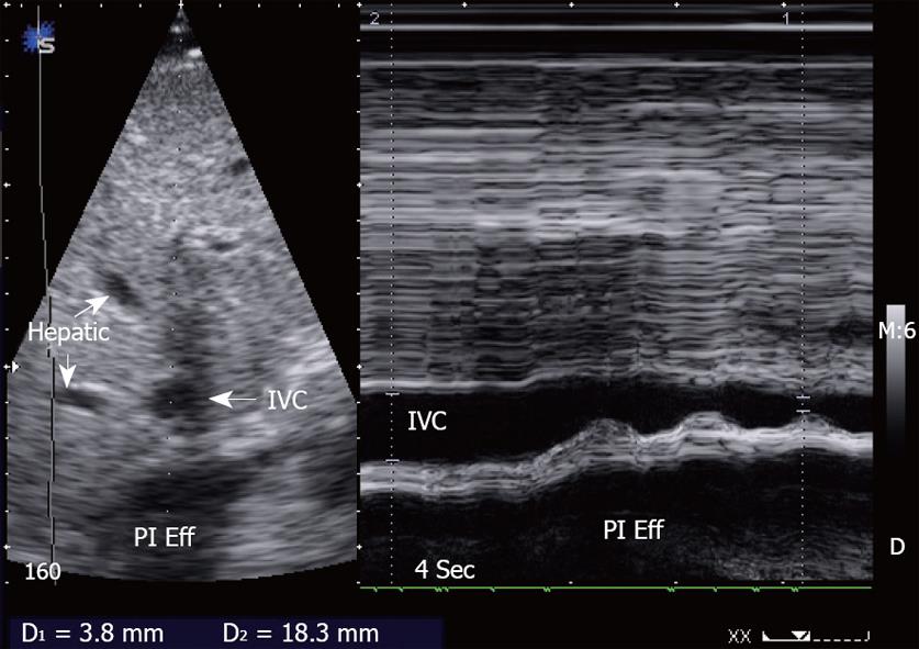
Figure 3 Inferior vena cava collapsibility index can be obtained from measurements of end-expiratory and end-inspiratory vena cava diameters.
The inferior vena cava collapsibility index (IVCCI) can be derived by subtracting the end-expiratory IVC diameter (IVCexp) from end-inspiratory IVC diameter (IVCinsp), and the dividing the difference by IVCinsp. The resultant fraction is then multiplied by 100% to produce a percentage IVC collapse. In this example, the IVCCI is [(18.3-3.8)/18.3] × 100% = 79.2%, which indicates hypovolemia. Pl Eff: Pleural effusion.
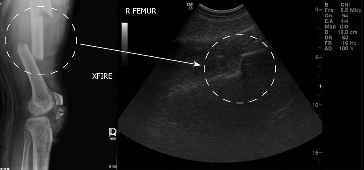
Figure 4 Bedside sonographic appearance of a displaced femoral fracture.
A correlation to plain radiography is shown. Bedside sonography provides a viable tool for quick and reliable assessment of suspected long-bone fractures. R FEMUR: Right femur; XFIRE: Cross table film.
- Citation: Stawicki SP, Howard JM, Pryor JP, Bahner DP, Whitmill ML, Dean AJ. Portable ultrasonography in mass casualty incidents: The CAVEAT examination. World J Orthop 2010; 1(1): 10-19
- URL: https://www.wjgnet.com/2218-5836/full/v1/i1/10.htm
- DOI: https://dx.doi.org/10.5312/wjo.v1.i1.10


