Copyright
©2010 Baishideng Publishing Group Co.
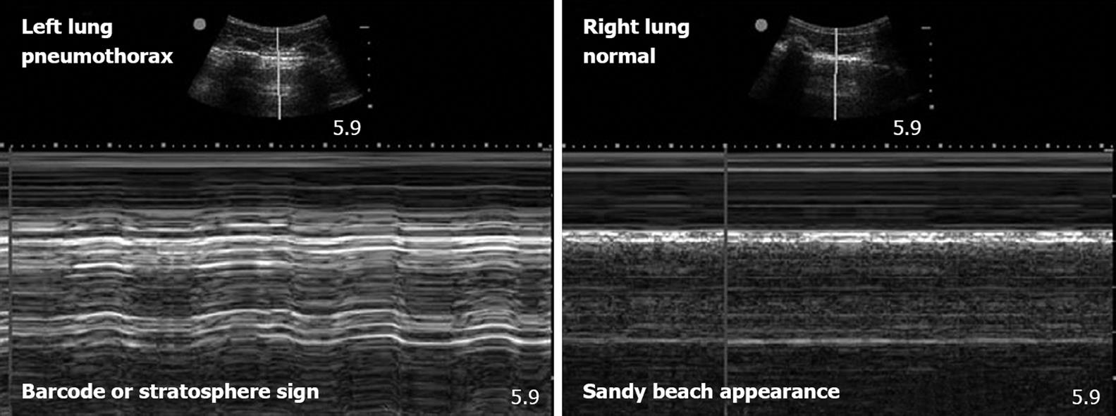
Figure 1 Pneumothorax on bedside ultrasonography.
These two images utilize M-mode echography to gather more information on the pleural interfaces seen as a hyperechoic line in the B-mode images above. The M-mode tracings below represent a single line of sight through the B-mode images. Over time, the gray-scale appearance of M-mode images changes with the movement of the tissue. In normal lungs the pleurae slide across each other and this movement takes on a “granular” or “sandy beach” appearance (image on the right). As a result of the lack of pleural sliding in the presence of pneumothorax, a pattern of horizontal striations that do not demonstrate the granular appearance of movement is present. This M-mode pattern that is characteristic of pneumothorax has been described as a “barcode” or the “stratosphere” sign (image on the left).
- Citation: Stawicki SP, Howard JM, Pryor JP, Bahner DP, Whitmill ML, Dean AJ. Portable ultrasonography in mass casualty incidents: The CAVEAT examination. World J Orthop 2010; 1(1): 10-19
- URL: https://www.wjgnet.com/2218-5836/full/v1/i1/10.htm
- DOI: https://dx.doi.org/10.5312/wjo.v1.i1.10


