
















Copyright: ©Author(s) 2026.
World J Clin Oncol. Sep 24, 2026; 17(9): 122832
Published online Sep 24, 2026. doi: 10.5306/wjco.122832
Published online Sep 24, 2026. doi: 10.5306/wjco.122832
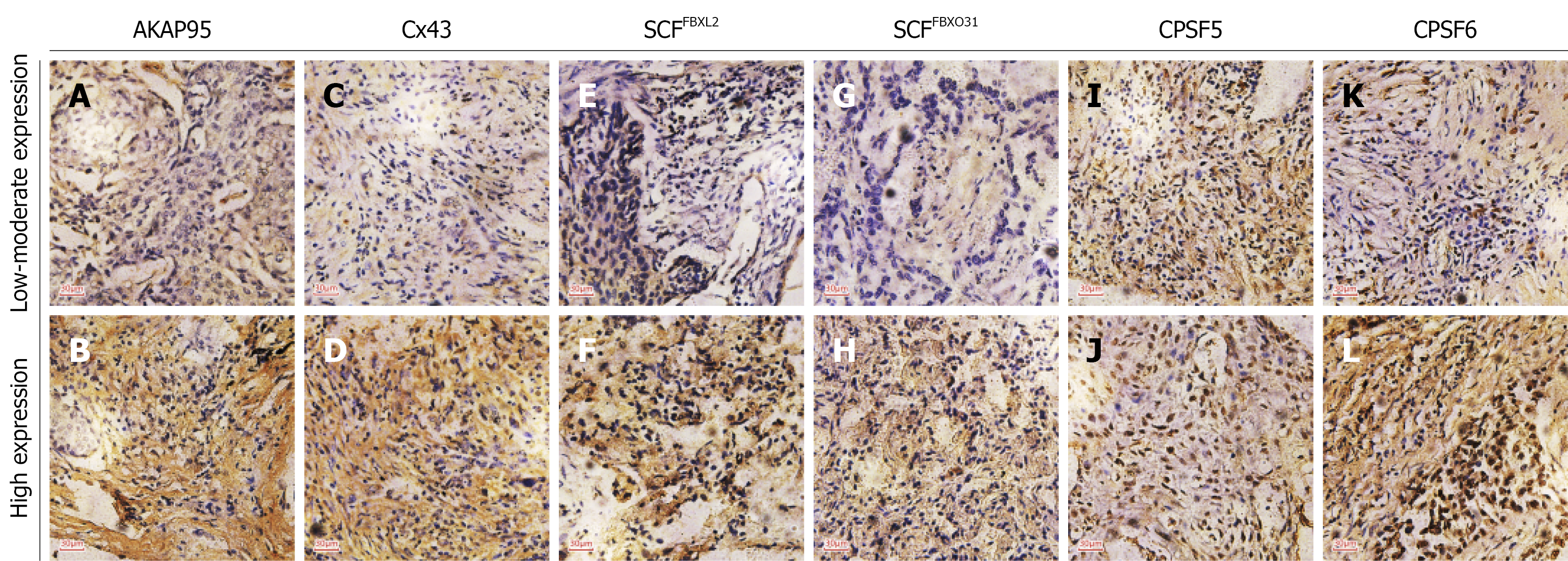
Figure 1 Expression of AKAP95, Cx43, SCFFBXL2, SCFFBXO31 CPSF5, and CPSF6 in lung cancer tissue cases.
A and B: Low and high expression of AKAP95, respectively, in both the cytoplasm and nucleus; C and D: Low and high expression of Cx43, respectively, in both the cytoplasm and nucleus; E and F: Low and high expression of SCFFBXL2, respectively, predominantly in the cytoplasm; G and H: Low and high expression of SCFFBXO31, respectively, predominantly in the cytoplasm; I and J: Moderate and high expression of CPSF5, respectively, primarily in the nucleus; K and L: Low and high expression of CPSF6, respectively, primarily in the nucleus. Scale bars: 30 μm.
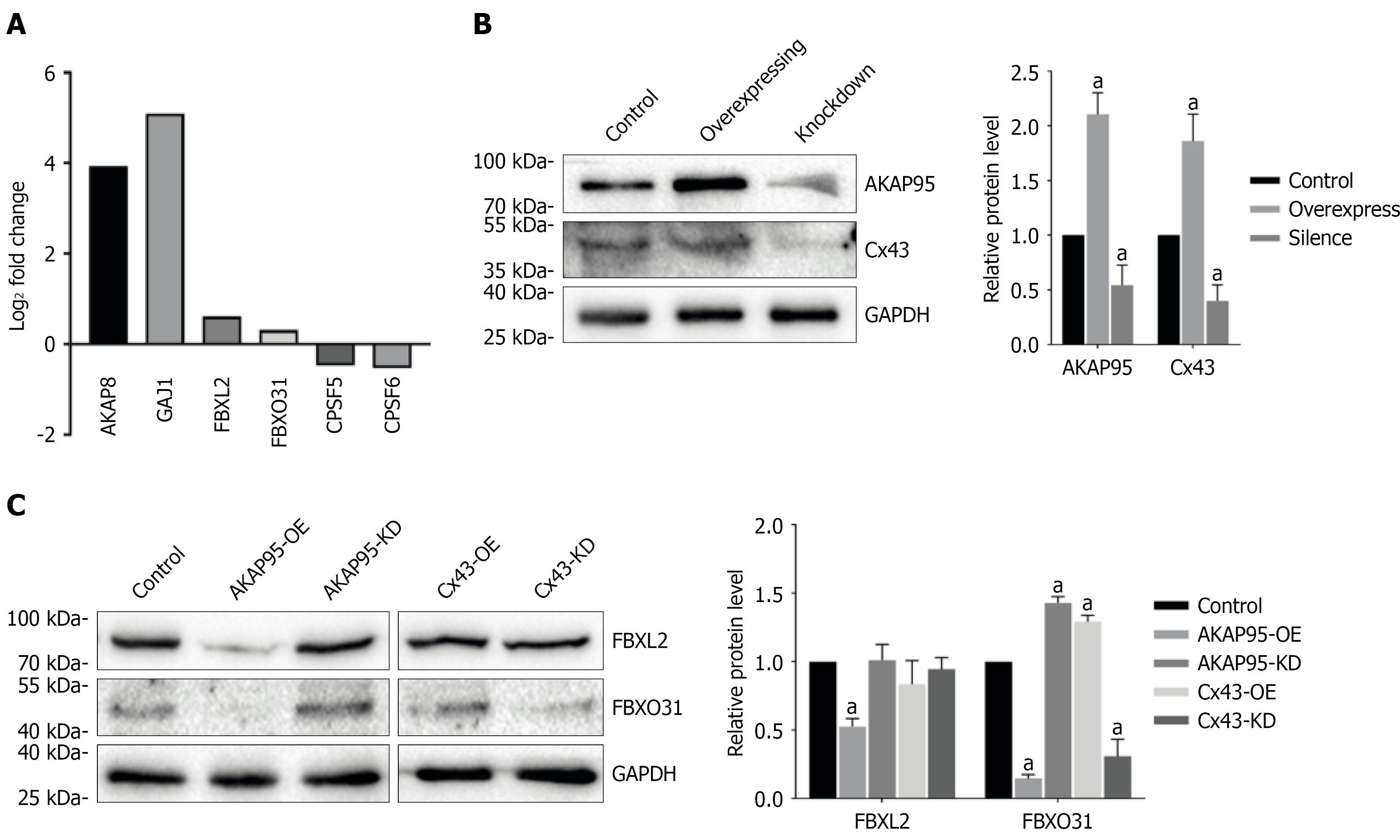
Figure 2 Expressions of SCFFBXL2 and SCFFBXO31 in AKAP95/Cx43 in A549 cells following AKAP95 or Cx43 overexpression or knockdown.
A: RNA-sequencing results from AKAP95-overexpressing cells showing increased mRNA expression levels of Cx43, SCFFBXL2, and SCFFBXO31; B: Decreased transcription levels of CPSF5 and CPSF6; C: Protein expression of AKAP95 and Cx43 in control, overexpression, and knockdown cells. Expressions of SCFFBXL2 and SCFFBXO31 following AKAP95 or Cx43 overexpression or knockdown in A549 cells. SCFFBXL2 expression decreased only in AKAP95-overexpressing cells. SCFFBXO31 expression was increased following Cx43 overexpression or AKAP95 knockdown and was decreased following AKAP95 overexpression or Cx43 knockdown. Band intensities were quantified by densitometry and normalized to GAPDH, with the corresponding matched control value set to 1 for each independent experiment. Data are presented as the mean ± SD of three independent experiments. aP < 0.05, indicates a statistically significant difference relative to the corresponding control.
- Citation: Deng ZF, Zhao L, Yuan Y, Li LK, Ge MY, Dou LD, Zhang JW, Wang D, Guo DB, An R, Zhang YX, Song B. Correlations of the expression of AKAP95, Cx43, CPSF5/6, SCFFBXL2 and SCFFBXO31 in lung cancer tissues. World J Clin Oncol 2026; 17(9): 122832
- URL: https://www.wjgnet.com/2218-4333/full/v17/i9/122832.htm
- DOI: https://dx.doi.org/10.5306/wjco.122832