Copyright
©The Author(s) 2025.
World J Clin Oncol. Aug 24, 2025; 16(8): 108557
Published online Aug 24, 2025. doi: 10.5306/wjco.v16.i8.108557
Published online Aug 24, 2025. doi: 10.5306/wjco.v16.i8.108557
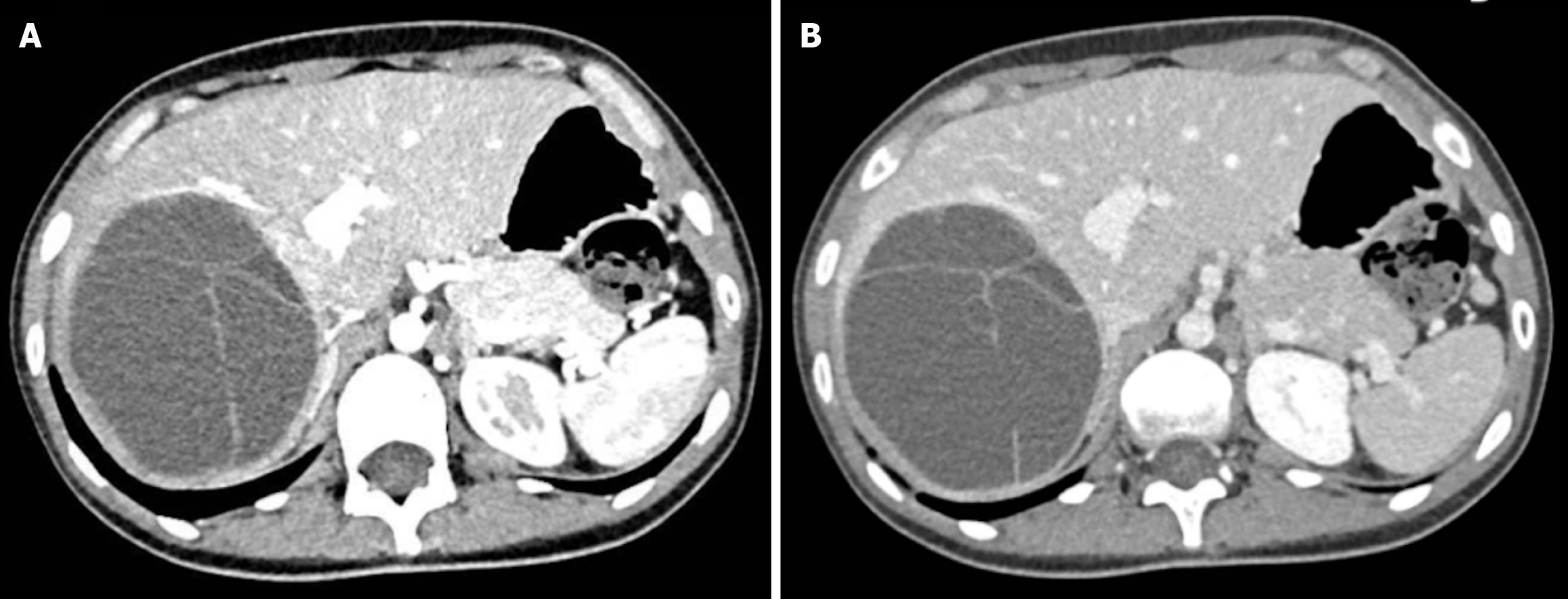
Figure 1 Contrast-enhanced computed tomography scan.
A and B: Demonstrating arterial (A) and venous (B) phases of a mucinous cystic neoplasm of the liver characterized by multiple enhancing septations.
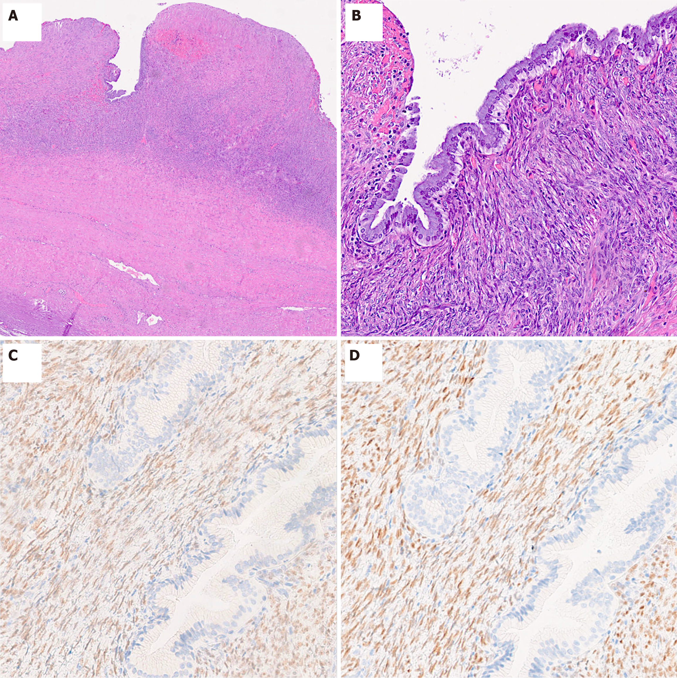
Figure 2 Histological aspect of a mucinous cystic neoplasms of the liver.
A: Low-power view of the cystic wall showing an extensively eroded epithelium and underlying ovarian-like stroma (HE 2.5×); B: Cyst lining predominantly composed of mucinous epithelium, overlying a hypercellular ovarian-like subepithelial stroma (HE 15×); C: Immunohistochemical staining showing estrogen receptor expression within the stromal component (15×); D: Immunohistochemical staining showing progesterone receptor expression within the stromal component (15×).
- Citation: Cicerone O, Basilico G, Tassi C, Antoniacomi C, Lucev F, Corallo S, Vanoli A, Maestri M. Mucinous cystic neoplasms of the liver: Literature review and case series. World J Clin Oncol 2025; 16(8): 108557
- URL: https://www.wjgnet.com/2218-4333/full/v16/i8/108557.htm
- DOI: https://dx.doi.org/10.5306/wjco.v16.i8.108557


