Copyright
©The Author(s) 2021.
World J Clin Oncol. Sep 24, 2021; 12(9): 823-832
Published online Sep 24, 2021. doi: 10.5306/wjco.v12.i9.823
Published online Sep 24, 2021. doi: 10.5306/wjco.v12.i9.823
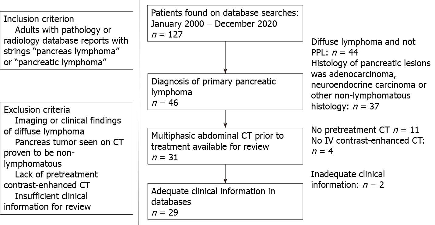
Figure 1 Inclusion and exclusion criteria of primary pancreatic lymphoma cohort.
This figure gives the derivation of our cohort of 29 subjects for this study. CT: Computed tomography; PPL: Primary pancreatic lymphoma.
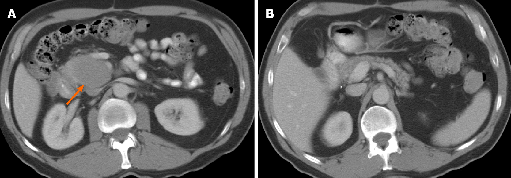
Figure 2 A 58-year-old male with abdominal pain.
A: Axial computed tomography images demonstrate a large, relatively well-defined focal mass (arrow) involving the uncinate process of the pancreas; B: Axial computed tomography images. The tumor shows homogenous hypoenhancement, without evidence of any necrosis or calcification. The rest of the pancreas is preserved, and there is no pancreatic ductal dilation. Biopsy confirmed diagnosis of primary pancreatic lymphoma.
Figure 3 A 72-year-old male presenting with abdominal pain and jaundice.
Coronal computed tomography imaging shows hypoenhancing homogenous infiltrative mass (thick arrows, A) infiltrating the mesenteric root. A: There is pancreatic duct dilation (short thin arrow) and common bile duct obstruction (arrowhead); B: There is stenosis of the superior mesenteric vein (long thin arrow) caused by the tumor.
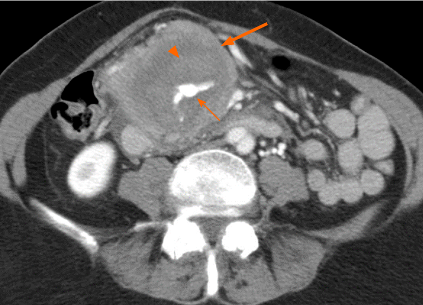
Figure 4 A 74-year-old female presenting with abdominal pain and nausea.
Axial computed tomography shows mass (thick arrow) encircling an aneurysmal vessel (thin arrow) which is bleeding into the mass, causing hematoma (arrowhead). The tumor was biopsy-proven to be primary pancreatic lymphoma.
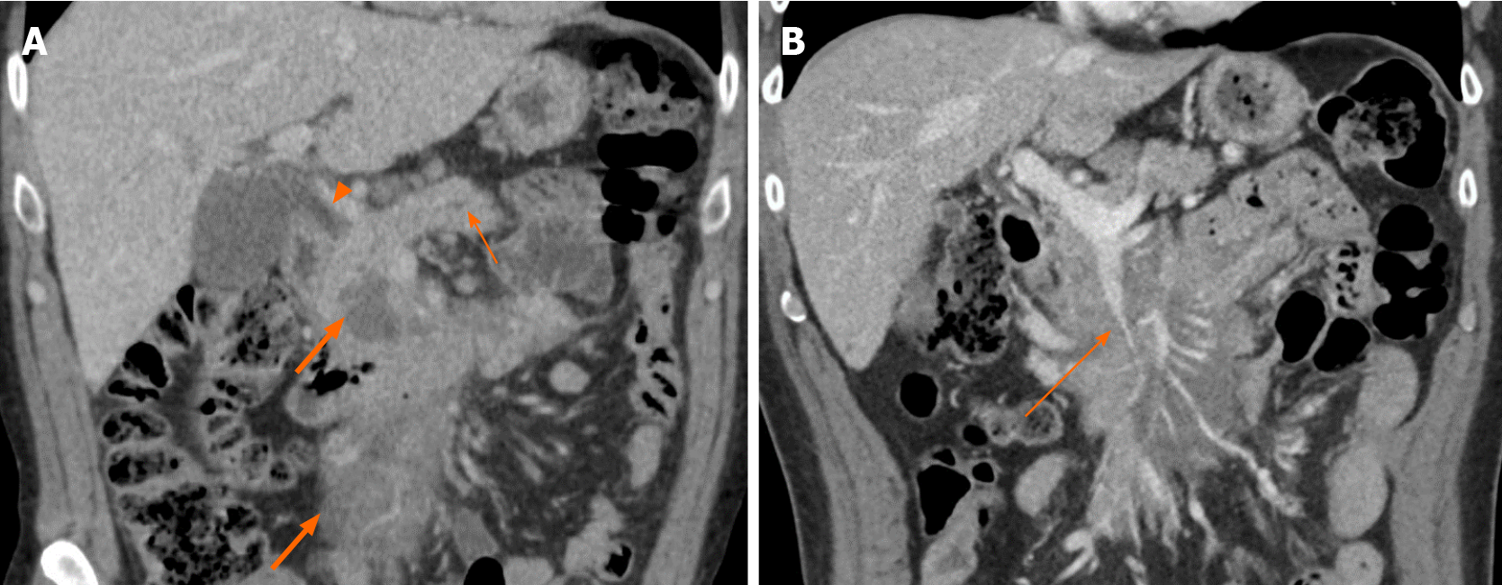
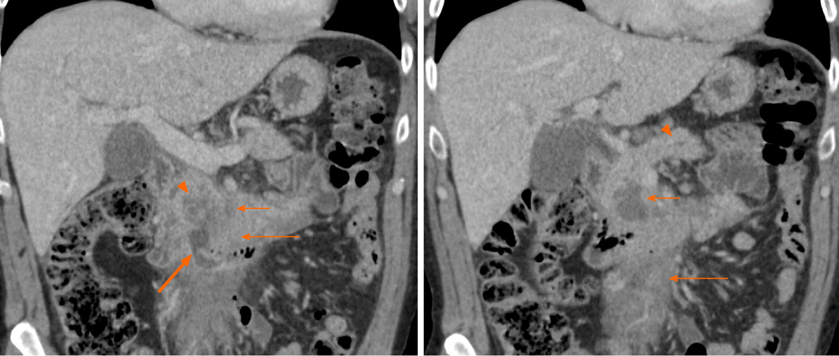
Figure 5 A 62-year-old male presenting with abdominal pain.
Coronal computed tomography images show low-density necrosis (thin short arrow) within tumor (thin long arrow), which infiltrates the mesenteric root without occlusion of mesenteric vessels. There is a collection extending from the third part of the duodenum into tumor (thick arrow) suggesting fistula from probable erosion of duodenal wall. Mild pancreatic duct dilation is seen (arrowhead).
- Citation: Segaran N, Sandrasegaran K, Devine C, Wang MX, Shah C, Ganeshan D. Features of primary pancreatic lymphoma: A bi-institutional review with an emphasis on typical and atypical imaging features. World J Clin Oncol 2021; 12(9): 823-832
- URL: https://www.wjgnet.com/2218-4333/full/v12/i9/823.htm
- DOI: https://dx.doi.org/10.5306/wjco.v12.i9.823


