
















©The Author(s) 2021.
World J Clin Oncol. Sep 24, 2021; 12(9): 823-832
Published online Sep 24, 2021. doi: 10.5306/wjco.v12.i9.823
Published online Sep 24, 2021. doi: 10.5306/wjco.v12.i9.823
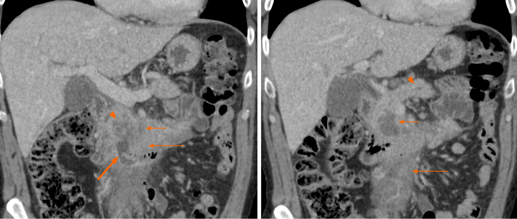
Figure 5 A 62-year-old male presenting with abdominal pain.
Coronal computed tomography images show low-density necrosis (thin short arrow) within tumor (thin long arrow), which infiltrates the mesenteric root without occlusion of mesenteric vessels. There is a collection extending from the third part of the duodenum into tumor (thick arrow) suggesting fistula from probable erosion of duodenal wall. Mild pancreatic duct dilation is seen (arrowhead).
- Citation: Segaran N, Sandrasegaran K, Devine C, Wang MX, Shah C, Ganeshan D. Features of primary pancreatic lymphoma: A bi-institutional review with an emphasis on typical and atypical imaging features. World J Clin Oncol 2021; 12(9): 823-832
- URL: https://www.wjgnet.com/2218-4333/full/v12/i9/823.htm
- DOI: https://dx.doi.org/10.5306/wjco.v12.i9.823