Copyright
©2010 Baishideng Publishing Group Co.
World J Gastrointest Pharmacol Ther. Oct 6, 2010; 1(5): 119-122
Published online Oct 6, 2010. doi: 10.4292/wjgpt.v1.i5.119
Published online Oct 6, 2010. doi: 10.4292/wjgpt.v1.i5.119
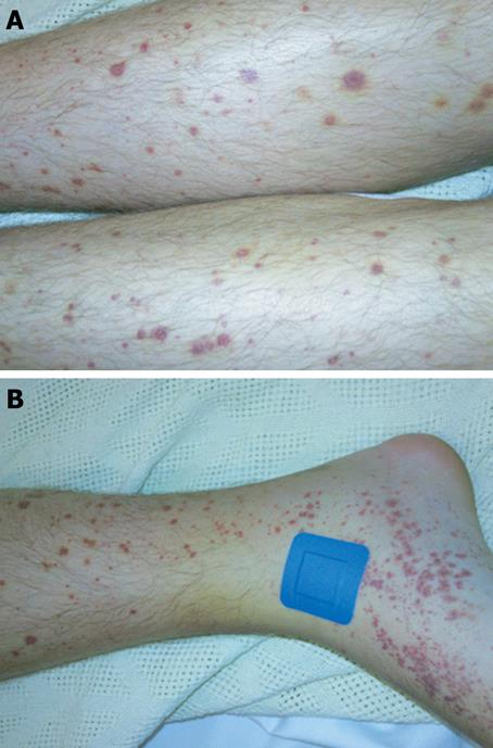
Figure 1 Vasculitic skin rash of Henoch-Schönlein purpura.
A: Typical palpable purpuric lesions on the legs; B: Site of skin punch biopsy.
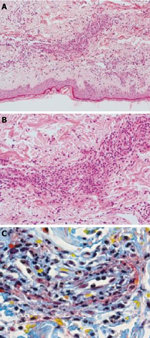
Figure 2 Punch biopsy of skin showing leukocytoclastic vasculitis.
A: Low (× 20); B: High (× 100) power views (H&E); C: MSB stain highlighting fibrinoid necrosis (red) and extravasation of red blood cells (yellow) in leukocytoclastic vasculitis.
- Citation: Rahman FZ, Takhar GK, Roy O, Shepherd A, Bloom SL, McCartney SA. Henoch-Schönlein purpura complicating adalimumab therapy for Crohn’s disease. World J Gastrointest Pharmacol Ther 2010; 1(5): 119-122
- URL: https://www.wjgnet.com/2150-5349/full/v1/i5/119.htm
- DOI: https://dx.doi.org/10.4292/wjgpt.v1.i5.119


