Published online Dec 28, 2016. doi: 10.4329/wjr.v8.i12.922
Peer-review started: April 12, 2016
First decision: May 19, 2016
Revised: August 3, 2016
Accepted: October 17, 2016
Article in press: October 18, 2016
Published online: December 28, 2016
Processing time: 253 Days and 8.6 Hours
To show imaging results from application of four-dimensional (4D) ultrasound lightening technique (HDlive™) in clinical obstetrics practice.
Normal and abnormal fetuses at second and third trimester of pregnancy undergoing routine scan with 4D HDlive™ (5DUS) in the rendering mode are described. Realistic features of fetal structures were provided by 5DUS in the rendering mode. Normal anatomy as well as pathology like cleft lip, hypoplastic face, micrognathia, low-set ears, corpus callosum, arthrogryposis, aortic arch, left congenital diaphragmatic hernia are highlighted in this study. Anatomical details of the fetuses were provided by 5DUS with higher quality imaging modality compared to those obtained using conventional 2D/3D ultrasound.
Realistic views of fetal anatomy details were displayed by means of 5DUS in the rendering mode, with high image quality obtained either in low-risk or in high-risk obstetrics population. Corpus callosum, esophagus, and aortic arch were obtained in normal fetuses. Cleft lip, cleft lip and palate, micrognathia, hypoplastic face, low-set ears, arthrogryposis, left congenital diaphragmatic hernia, exomphalos, and clitoris hypertrophy were clearly rendered by 5DUS application.
The use of 5DUS in the rendering mode, when clinical available, was diagnostic in a variety of congenital anomalies, aided understanding of the parents-to-be and improved prenatal counseling and perinatal management.
Core tip: Four-dimensional ultrasound using HDlive™ allows realistic images of fetal anatomic structures in the second trimester of pregnancy. These images allow identifying fine details of fetal surface, with better understanding both multidisciplinary team and parents.
- Citation: Tonni G, Grisolia G, Santana EF, Júnior EA. Assessment of fetus during second trimester ultrasonography using HDlive software: What is its real application in the obstetrics clinical practice? World J Radiol 2016; 8(12): 922-927
- URL: https://www.wjgnet.com/1949-8470/full/v8/i12/922.htm
- DOI: https://dx.doi.org/10.4329/wjr.v8.i12.922
The second trimester scan, also called anomaly scan, is usually performed between 18-23 wk, and is based on a systematic anatomical survey of the fetus, placenta and umbilical cord, in order to detect possible fetal abnormalities[1]. The ultrasound examination should be carried out according to international standard[2] and possibly by accredited sonographers who have completed appropriate training program by scientific societies[3].
The sensitivity and specificity for detection of congenital anomalies by means of conventional 2D ultrasound may be estimated around 83.5% and 99.8%, respectively[4,5]. The technologic advancement gained by real-time, high definition three- and four-dimensional ultrasound (3D/4DUS), enable acquisition of volume that can be analysed online or offline by “navigating” within the volume in the three orthogonal planes. 3D/4DUS post-processing techniques allow anatomical details to be investigated in sagittal, axial and coronal planes, improving prenatal diagnosis of congenital malformations[6-8].
Hereafter, we present a pictorial editorial from normal and pathologic cases obtained during second and third trimester of pregnancy in low- and in high-risk pregnancy using 4D HDlive™ (5DUS) software.
Ultrasound examinations were performed using Voluson E8 apparatus equipped with a transabdominal volumetric RAB4D ultrasound probe (GE, Milwaukee, WI). Fetal anatomical survey was performed using conventional 2D ultrasound, and 3D/4D HDlive™ (5DUS) applied both in low- and in high-risk pregnancy. The study was approved by the local Ethics Committee of both Guastalla Civil (AUSL Reggio Emilia) and “Carlo Poma” hospitals (AUSL Mantua), Italy. Four-hundred low-risk and seventy-six high-risk pregnant women entering the clinical trial gave written informed consent. Two consecutive volumes were acquired during transient maternal apnea and fetal rest to reduce motion artefacts. The sweep took less just than few seconds. Acquisition angle of 45-60 degree was used, depending on the gestational age. All 3DUS volumes were saved both onto the ultrasound equipment and onto a optical disk for post-processing analysis. 5DUS application was applied to the best 3DUS volume stored and different lightening and shadowing adjustments were made to obtain the highest image quality rendering. Offline analysis was performed using a computer developed platform (4DView™, Zipf, Austria); HDlive™ (5DUS) software was applied after uploading the software onto a personal computer using a freely released flash-drive pen.
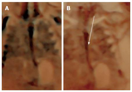
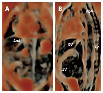
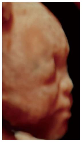
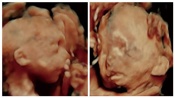
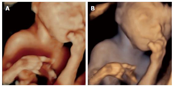
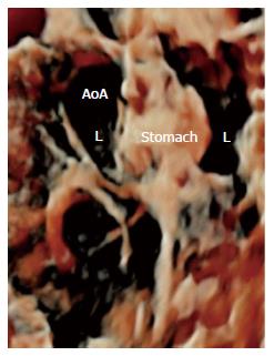
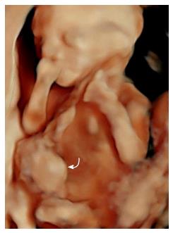
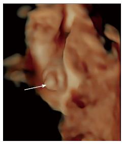
Realistic views of fetal anatomy details were displayed by means of 5DUS in the rendering mode, with high image quality obtained either in low-risk or in high-risk obstetrics population. Corpus callosum (Figure 1), esophagus (Figure 2), and aortic arch (Figure 3) were obtained in normal fetuses.
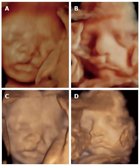
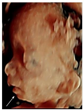
Cleft lip (Figure 4), cleft lip and palate (Figure 5), micrognathia (Figure 6), hypoplastic face (Figure 7), low-set ears (Figure 8), arthrogryposis (Figure 9), left congenital diaphragmatic hernia (Figure 10), exomphalos (Figure 11), and clitoris hypertrophy (Figure 12) were clearly rendered by 5DUS application.
This pictorial editorial displays a gallery of normal and pathologic cases obtained during second and third trimester of pregnancy by means of 5DUS in the rendering mode. 3D/4DUS with its technical applications has resulted in improved diagnostic accuracy compared with conventional 2DUS, especially when applied to the field of fetal medicine, where high definition 3D/4DUS produces real-time reconstruction of the fetal anatomy[9-13]. HDlive™ imaging often looks like a picture taken inside the uterus[14] and may enable detection of subtle malformations that may go undiagnosed using conventional 2DUS. This may be particularly seen when dealing with surface abnormalities such as those involving the fetal face where 4DUS, especially with HDlive™ rendering mode, may offers a potential imaging enhancement. Previous observation has documented a role for 3DUS to provide additional information compared to 2DUS for the prenatal diagnosis of facial, skeletal and neural tube defect[15]. An extended review from Tonni et al[16] has described the technical advancements obtained over the past 20 years by 3D/4DUS compared to conventional 2DUS in different fields of application, particularly in prenatal diagnosis. The study of the fetal face, palate and detection rate of cleft lip and cleft palates has resulted enhanced when 3D/4DUS has complemented 2DUS, either in the first as in the second trimester of pregnancy[17-23]. Undoubtedly, one of the main advantages of 3D/4DUS is represented by the possibility of volume acquisition compared with “flat” images obtained by 2DUS. Once a volume is acquired, it can be further manipulate by “navigating” online or offline within the volume. In addition, anatomical details can be displayed in all the three orthogonal planes. Furthermore, 3D/4DUS can be used in training program as the volume can be freely section on demand and send to expert at remote site using DICOM (digital communication in medicine) technology[16]. Moreover, observations have shown that 4DUS has been a valuable diagnostic investigation to assess fetal neurobehavioral state as it allows visualization of yawning, sucking, smiling, and blinking activity[13,14]. 5DUS differs from conventional rendering methods because it uses a fixed virtual light source that calculates the propagation of light through skin and tissue. Operators can freely select the light source at any angle relative to the ultrasound volume to enhance anatomical details[11]. 5DUS is a relatively easy technique to be applied and does not require specific clinical training for operators already confident with post-processing 3D/4DUS techniques. The time needed to obtain online the desired reconstructed image and display it on the ultrasound screen can be estimated usually in about 1 minute, depending upon experience gained and anatomical details that need to be rendered. For comparison analysis of image quality between 3DUS vs 4D HDlive™, it is advisable to save and stored the 3DUS volumes on the ultrasound apparatus and to export them onto a flash-drive pen or onto an optical disk for further offline post-processing analysis. This is required because once HDlive™ is applied to a 3DUS volume, the rendered image will be saved automatically in this modality and previous 3DUS volume is lost. 5DUS may represents a complementary diagnostic tool to confirm fetal abnormalities and to characterize anatomical details such as those seen in rare syndromes thus improving accurate prenatal diagnosis, genetic counseling and antenatal management in targeted cases. Importantly, these “life-like” images provided by HDlive™ may represent a technological improvement in 3D imaging that may strengthen the maternal-fetal bonding process[24,25]. HDlive™ software has shown limitations in conditions of poor imaging quality, such as in cases of increased maternal body mass index, presence of abdominal scar or uterine myomata as well as fetal positioning in utero. In the current clinical trial, unsuccessful volume acquisition for adequate 4D HDlive™ rendering has occurred in 6.75% of cases in low-risk and 3.9% in high-risk pregnancies. However, some of these clinical limitations may be overwhelm by transvaginal approach. Nonetheless, further studies will be needed to assess the role of 5DUS and its clinical validation before the use of this advanced lightening software may be included in obstetrics practice and be used at the time of routine scan in low-risk women or applied to the study of structural fetal malformations in high-risk pregnancies.
The second trimester scan, also called anomaly scan, is usually performed between 18-23 wk, and is based on a systematic anatomical survey of the fetus, placenta and umbilical cord, in order to detect possible fetal abnormalities. The ultrasound examination should be carried out according to international standard and possibly by accredited sonographers who have completed appropriate training program by scientific societies. The sensitivity and specificity for detection of congenital anomalies by means of conventional 2D ultrasound may be estimated around 83.5% and 99.8%, respectively.
The technologic advancement gained by real-time, high definition three- and four-dimensional ultrasound (3D/4DUS), enable acquisition of volume that can be analysed online or offline by “navigating” within the volume in the three orthogonal planes. Previous observation has documented a role for 3DUS to provide additional information compared to 2DUS for the prenatal diagnosis of facial, skeletal and neural tube defect.
The authors present a pictorial editorial from normal and pathologic cases obtained during second and third trimester of pregnancy in low- and in high-risk pregnancy using 4D HDliveTM (5DUS) software.
3D/4DUS post-processing techniques allow anatomical details to be investigated in sagittal, axial and coronal planes, improving prenatal diagnosis of congenital malformations.
Well written manuscript, nice pictures.
Manuscript source: Invited manuscript
Specialty type: Radiology, nuclear medicine and medical imaging
Country of origin: Brazil
Peer-review report classification
Grade A (Excellent): A
Grade B (Very good): 0
Grade C (Good): C
Grade D (Fair): 0
Grade E (Poor): 0
P- Reviewer: Rizzo G, Rovas L S- Editor: Kong JX L- Editor: A E- Editor: Wu HL
| 1. | Pilu G, Nicolaides KH, Diagnosis of fetal abnormalities. The 18-23 scan. 1st ed. London: Parthenon Publishing 1999; . |
| 2. | Salomon LJ, Alfirevic Z, Berghella V, Bilardo C, Hernandez-Andrade E, Johnsen SL, Kalache K, Leung KY, Malinger G, Munoz H. Practice guidelines for performance of the routine mid-trimester fetal ultrasound scan. Ultrasound Obstet Gynecol. 2011;37:116-126. [RCA] [PubMed] [DOI] [Full Text] [Cited by in Crossref: 646] [Cited by in RCA: 640] [Article Influence: 45.7] [Reference Citation Analysis (0)] |
| 3. | International Society of Ultrasound in Obstetrics & Gynecology Education Committee. Sonographic examination of the fetal central nervous system: guidelines for performing the ‘basic examination’ and the “fetal neurosonogram”. Ultrasound Obstet Gynecol. 2007;29:109-116. [RCA] [PubMed] [DOI] [Full Text] [Cited by in Crossref: 450] [Cited by in RCA: 397] [Article Influence: 22.1] [Reference Citation Analysis (0)] |
| 4. | Grandjean H, Larroque D, Levi S. The performance of routine ultrasonographic screening of pregnancies in the Eurofetus Study. Am J Obstet Gynecol. 1999;181:446-454. [RCA] [PubMed] [DOI] [Full Text] [Cited by in Crossref: 370] [Cited by in RCA: 325] [Article Influence: 12.5] [Reference Citation Analysis (0)] |
| 5. | Bernaschek G, Stuempflen I, Deutinger J. The value of sonographic diagnosis of fetal malformations: different results between indication-based and screening-based investigations. Prenat Diagn. 1994;14:807-812. [RCA] [PubMed] [DOI] [Full Text] [Cited by in Crossref: 13] [Cited by in RCA: 13] [Article Influence: 0.4] [Reference Citation Analysis (0)] |
| 6. | Tonni G, Araujo Júnior E. Three-dimensional ultrasound in obstetrics practice: myth or reality? Rev Bras Ginecol Obstet. 2014;36:143-145. [RCA] [PubMed] [DOI] [Full Text] [Cited by in Crossref: 2] [Cited by in RCA: 2] [Article Influence: 0.2] [Reference Citation Analysis (0)] |
| 7. | Tonni G, Lituania M. OmniView algorithm: a novel 3-dimensional sonographic technique in the study of the fetal hard and soft palates. J Ultrasound Med. 2012;31:313-318. [PubMed] |
| 8. | Tonni G, Grisolia G, Sepulveda W. Second trimester fetal neurosonography: reconstructing cerebral midline anatomy and anomalies using a novel three-dimensional ultrasound technique. Prenat Diagn. 2014;34:75-83. [RCA] [PubMed] [DOI] [Full Text] [Cited by in Crossref: 24] [Cited by in RCA: 26] [Article Influence: 2.2] [Reference Citation Analysis (0)] |
| 9. | Hata T, Hanaoka U, Tenkumo C, Sato M, Tanaka H, Ishimura M. Three- and four-dimensional HDlive rendering images of normal and abnormal fetuses: pictorial essay. Arch Gynecol Obstet. 2012;286:1431-1435. [RCA] [PubMed] [DOI] [Full Text] [Cited by in Crossref: 63] [Cited by in RCA: 64] [Article Influence: 4.9] [Reference Citation Analysis (0)] |
| 10. | Santana EF, Helfer TM, Piassi Passos J, Araujo Júnior E. Prenatal diagnosis of a giant epignathus teratoma in the third trimester of pregnancy using three-dimensional ultrasound and magnetic resonance imaging. Case report. Med Ultrason. 2014;16:168-171. [PubMed] [DOI] [Full Text] |
| 11. | Grisolia G, Tonni G. Fetal echocardiography using HDlive. J Obstet Gynaecol Can. 2013;35:497-498. [RCA] [PubMed] [DOI] [Full Text] [Cited by in Crossref: 8] [Cited by in RCA: 8] [Article Influence: 0.7] [Reference Citation Analysis (0)] |
| 12. | Araujo Júnior E, Nardozza LM, Moron AF. Three-dimensional ultrasound STIC-HDlive rendering: new technique to assessing of fetal heart. Rev Bras Cir Cardiovasc. 2013;28:v-vii. [RCA] [PubMed] [DOI] [Full Text] [Full Text (PDF)] [Cited by in Crossref: 5] [Cited by in RCA: 6] [Article Influence: 0.5] [Reference Citation Analysis (0)] |
| 13. | Grigore M, Mareş A. The role of HDlive technology in improving the quality of obstetrical images. Med Ultrason. 2013;15:209-214. [RCA] [PubMed] [DOI] [Full Text] [Cited by in Crossref: 18] [Cited by in RCA: 8] [Article Influence: 0.7] [Reference Citation Analysis (0)] |
| 14. | Yigiter AB, Kavak ZN. Normal standards of fetal behavior assessed by four-dimensional sonography. J Matern Fetal Neonatal Med. 2006;19:707-721. [RCA] [PubMed] [DOI] [Full Text] [Cited by in Crossref: 75] [Cited by in RCA: 59] [Article Influence: 3.3] [Reference Citation Analysis (0)] |
| 15. | Gonçalves LF, Lee W, Espinoza J, Romero R. Three- and 4-dimensional ultrasound in obstetric practice: does it help? J Ultrasound Med. 2005;24:1599-1624. [PubMed] |
| 16. | Tonni G, Martins WP, Guimarães Filho H, Araujo Júnior E. Role of 3-D ultrasound in clinical obstetric practice: evolution over 20 years. Ultrasound Med Biol. 2015;41:1180-1211. [RCA] [PubMed] [DOI] [Full Text] [Cited by in Crossref: 26] [Cited by in RCA: 26] [Article Influence: 2.6] [Reference Citation Analysis (0)] |
| 17. | Tonni G, Grisolia G, Sepulveda W. Early prenatal diagnosis of orofacial clefts: evaluation of the retronasal triangle using a new three-dimensional reslicing technique. Fetal Diagn Ther. 2013;34:31-37. [RCA] [PubMed] [DOI] [Full Text] [Cited by in Crossref: 24] [Cited by in RCA: 24] [Article Influence: 2.0] [Reference Citation Analysis (0)] |
| 18. | Tonni G, Rosignoli L, Palmisano M, Sepulveda W. Early Detection of Cleft Lip by Three-Dimensional Transvaginal Ultrasound in Niche Mode in a Fetus With Trisomy 18 Diagnosed by Celocentesis. Cleft Palate Craniofac J. 2016;53:745-748. [RCA] [PubMed] [DOI] [Full Text] [Cited by in Crossref: 6] [Cited by in RCA: 6] [Article Influence: 0.6] [Reference Citation Analysis (0)] |
| 19. | Tonni G, Grisolia G. Fetal uvula: navigating and lightening the soft palate using HDlive. Arch Gynecol Obstet. 2013;288:239-244. [RCA] [PubMed] [DOI] [Full Text] [Cited by in Crossref: 13] [Cited by in RCA: 9] [Article Influence: 0.8] [Reference Citation Analysis (0)] |
| 20. | Tonni G, Centini G, Rosignoli L. Prenatal screening for fetal face and clefting in a prospective study on low-risk population: can 3- and 4-dimensional ultrasound enhance visualization and detection rate? Oral Surg Oral Med Oral Pathol Oral Radiol Endod. 2005;100:420-426. [RCA] [PubMed] [DOI] [Full Text] [Cited by in Crossref: 37] [Cited by in RCA: 29] [Article Influence: 1.5] [Reference Citation Analysis (0)] |
| 21. | Sepulveda W, Cafici D, Bartholomew J, Wong AE, Martinez-Ten P. First-trimester assessment of the fetal palate: a novel application of the Volume NT algorithm. J Ultrasound Med. 2012;31:1443-1448. [PubMed] |
| 22. | Sepulveda W, Wong AE, Castro F, Adiego B, Martinez-Ten P. Feasibility of 3-dimensional sonographic examination of the fetal secondary palate during the second-trimester anatomy scan. J Ultrasound Med. 2011;30:1619-1624. [PubMed] |
| 23. | Martinez-Ten P, Adiego B, Illescas T, Bermejo C, Wong AE, Sepulveda W. First-trimester diagnosis of cleft lip and palate using three-dimensional ultrasound. Ultrasound Obstet Gynecol. 2012;40:40-46. [RCA] [PubMed] [DOI] [Full Text] [Cited by in Crossref: 47] [Cited by in RCA: 44] [Article Influence: 3.4] [Reference Citation Analysis (0)] |
| 24. | Ji EK, Pretorius DH, Newton R, Uyan K, Hull AD, Hollenbach K, Nelson TR. Effects of ultrasound on maternal-fetal bonding: a comparison of two- and three-dimensional imaging. Ultrasound Obstet Gynecol. 2005;25:473-477. [RCA] [PubMed] [DOI] [Full Text] [Cited by in Crossref: 75] [Cited by in RCA: 62] [Article Influence: 3.1] [Reference Citation Analysis (0)] |
| 25. | Rustico MA, Mastromatteo C, Grigio M, Maggioni C, Gregori D, Nicolini U. Two-dimensional vs. two- plus four-dimensional ultrasound in pregnancy and the effect on maternal emotional status: a randomized study. Ultrasound Obstet Gynecol. 2005;25:468-472. [RCA] [PubMed] [DOI] [Full Text] [Cited by in Crossref: 58] [Cited by in RCA: 49] [Article Influence: 2.5] [Reference Citation Analysis (0)] |


