Copyright
©2014 Baishideng Publishing Group Inc.
World J Radiol. Jul 28, 2014; 6(7): 511-514
Published online Jul 28, 2014. doi: 10.4329/wjr.v6.i7.511
Published online Jul 28, 2014. doi: 10.4329/wjr.v6.i7.511
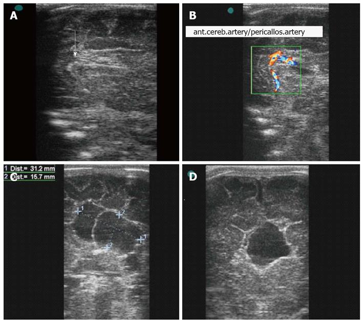
Figure 1 Transfontanellar ultrasonography.
A: In the sagittal plane, showing the knee of the corpus callosum (with arrow); the spleen and posterior segment were not observed; B: In the sagittal plane, showing partial agenesis of the corpus callosum with the aid of color Doppler on the pericallosal artery; C: In the parasagittal plane, showing a cyst in the inter-hemispherical fissure; D:In the coronal plane, showing a cyst in the inter-hemispherical fissure and porencephaly.
- Citation: Pires CR, Júnior EA, Czapkowski A, Zanforlin Filho SM. Aicardi syndrome: Neonatal diagnosis by means of transfontanellar ultrasound. World J Radiol 2014; 6(7): 511-514
- URL: https://www.wjgnet.com/1949-8470/full/v6/i7/511.htm
- DOI: https://dx.doi.org/10.4329/wjr.v6.i7.511


