Published online May 28, 2011. doi: 10.4329/wjr.v3.i5.135
Revised: March 24, 2011
Accepted: May 1, 2011
Published online: May 28, 2011
AIM: To investigate the features of abdominal crush injuries resulting from an earthquake using multidetector computed tomography (MDCT).
METHODS: Fifty-one survivors with abdominal crush injuries due to the 2008 Sichuan earthquake underwent emergency non-enhanced scans with 16-row MDCT. Data were reviewed focusing on anatomic regions including lumbar vertebrae, abdominal wall soft tissue, retroperitoneum and intraperitoneal space; and types of traumatic lesions.
RESULTS: Fractures of lumbar vertebrae and abdominal wall soft tissue injuries were more common than retro- and intraperitoneal injuries (P < 0.05). With regard to the 49 lumbar vertebral fractures in 24 patients, these occurred predominantly in the transverse process (P < 0.05), and 66.67% of patients (16/24) had fractures of multiple vertebrae, predominantly two vertebrae in 62.5% of patients (10/16), mainly in L1-3 vertebrae in 81.63% of the vertebrae (40/49). Retroperitoneal injuries occurred more frequently than intraperitoneal injuries (P < 0.05), and renal and liver injuries were most often seen in the retroperitoneum and in the intraperitoneal space, respectively (all P < 0.05).
CONCLUSION: Transverse process fractures in two vertebrae among L1-3 vertebrae, injury of abdominal wall soft tissue, and renal injury might be features of earthquake-related crush abdominal injury.
- Citation: Chen TW, Yang ZG, Dong ZH, Shao H, Chu ZG, Tang SS. Abdominal crush injury in the Sichuan earthquake evaluated by multidetector computed tomography. World J Radiol 2011; 3(5): 135-140
- URL: https://www.wjgnet.com/1949-8470/full/v3/i5/135.htm
- DOI: https://dx.doi.org/10.4329/wjr.v3.i5.135
An earthquake that registered 8.0 on the Richter scale devastated the mountainous region of Sichuan in China at 2:28 pm Beijing time on May 12, 2008, and the epifocus was located at Wenchuan County in the Sichuan Province of China. The widespread effect of the earthquake destroyed a significant number of schools, factories, apartments, office areas, and villages. The schools, factories and villages were more vulnerable compared with the other styles of buildings. Due to the high-force impacts of building collapse or falling objects in this earthquake, 374 643 people were injured with 69 227 killed and 17 923 missing. In view of the massive morbidity arising from this event, an undamaged key university hospital 92 kilometers away from the epicenter, equipped with 4300 beds, working as the largest-scale urgent care center in the earthquake-affected areas, received the largest number of injured people, and treated a total of 2728 cases over a period of 15 d. Of these patients, 2.05% (56/2728) had sustained abdominal injury associated with crush injury. Missed abdominal injury or delayed treatment frequently results in preventable mortality, but prompt localization of abdominal injuries in trauma patients can improve the efficacy of injury management[1,2].
To localize suspected blunt abdominal injury, ultrasound is used as an initial tool in most European and Asian-Pacific countries[3]. However, ultrasonography is not sensitive in detecting hollow organ and retroperitoneal injury[4]. Computed tomography (CT) scanning of the abdomen can overcome the limitation of ultrasonography, and is currently considered the gold standard for detecting intra- and retroperitoneal lesions in trauma patients[5-8]. To our knowledge, the features of abdominal crush injuries on CT due to massive earthquake damage have not been reported in the literature, although some relevant publications regarding crush injuries in other anatomic regions have been reported[9-12]. Thus, our aim was to retrospectively investigate the features of abdominal crush injuries resulting from this earthquake using emergency multidetector CT (MDCT) for better understanding and treatment planning of abdominal crush trauma survivors in similar future earthquakes.
The local institutional ethic review board approved the present study, and patient informed consent was waived. Patients entered our study according to the following inclusion criteria: (1) victims had abdominal crush injuries in combination with or without injuries in other systems such as the thorax and pelvis; (2) clinical suggested abdominal injuries were initially confirmed by CT; and (3) the etiology of abdominal injuries was crush injury in the 2008 Sichuan earthquake on the basis of the history and the findings of rescue. Patients with abdominal injuries were excluded from the present study if the etiology of the injuries was jumping or accidental falling from buildings based on the history of injury.
Between May 12 and May 26, 2008, fifty-one consecutive survivors (22 men and 29 women; mean age, 41.92 years; age range, 7-86 years) with abdominal crush injuries resulting from the 2008 Sichuan earthquake, who were admitted into this university hospital and met the inclusion criteria, were enrolled in the present study. In the cohort, 49 patients with abdominal crush injuries in combination with crush injuries in one or more other anatomic regions including the thorax, pelvis, extremity and neck were confirmed by MDCT scans in 39, 40, 11 and 4 patients, respectively. In addition, 36 patients including 27 patients with retro- or intraperitoneal visceral injuries had acute renal failure confirmed by the measurement of myoglobin in blood and urine shortly after the CT scans. In order to timely detect the injuries for appropriate emergency treatments in a great number of injured patients, two of five MDCT scanners in the university hospital were utilized to image the abdominal injuries as soon as possible. The mean time from injury to imaging was 5.4 d with a range of 6 h to 14 d. In patients who waited a long time from injury to rescue, few dangerously ill patients survived before being conveyed to the university hospital to receive effective treatments, although some had received antibiotics in the field hospital to prevent infection in the disaster areas. Based on the image findings and clinical data, 37 patients with retro- or intraperitoneal visceral injuries, fracture of lumber vertebra, or severe injury of abdominal wall soft tissue underwent surgery, and the remaining victims received conservative treatment. Owing to appropriate treatments, 49 patients were cured, and 2 patients died of fatal crush injury.
Thirty-eight and thirteen victims with abdominal crush injuries underwent CT scanning with a Philips Brilliance 16-row MDCT (Philips Healthcare, Eindhoven, the Netherlands) and a Siemens Somatom Sensation 16-row MDCT (Siemens Medical Systems, Forchheim, Germany), respectively. An emergency abdominal CT scan without intravenous contrast material was obtained as soon as possible from the right diaphragmatic dome to the pelvic basement. Because we suspected all victims to have abdominal injuries in combination with acute renal failure due to the massive earthquake, the patients did not undergo contrast-enhanced abdominal CT scans in the earthquake situation. The following scanning parameters were used for both scanners to image the injuries: 120 kV, 250 mAs, 0.5-s gantry rotation time, pitch of 0.85, collimation of 16 mm × 0.75 mm, 5-mm reconstructed section thickness, 380-mm field of view, and matrix of 512 mm × 512 mm. When fractures of the lumbar vertebrae were found, the reconstructed section thickness was 1 mm.
Image data were transferred to a picture archiving communication system (Syngo-Imaging, Siemens Medical Solutions, Forchheim, Germany). Data were retrospectively analyzed by an experienced radiological Associate Professor (the first author, who had 12 years of experience in radiology) and one experienced radiologist (the third author with 11 years of experience in radiology) working in consensus with emphasis on trauma in anatomic regions including the retroperitoneum, intraperitoneal space, lumbar vertebrae, and abdominal wall soft tissue. Because of lumbar vertebral fractures related to pathological changes in abdominal wall soft tissue, the fractures were reviewed in detail by multiplanar reconstruction with a slab of 5-7 mm and three-dimensional reconstruction. In order to better understand overall types of abdominal injuries, traumatic lesions in parenchymal organs together with peritoneal pathological changes (seroperitoneum and pneumoperitoneum) and injuries of abdominal aorta were reviewed.
Statistical analysis was performed with the SPSS statistical package (version 13.0 for Windows, SPSS Inc., Chicago, IL, USA). We compared incidences of lumbar vertebrae and abdominal wall soft tissue crush injuries with those of retro- and intraperitoneal visceral crush injuries using the Chi-square test. If significant differences were found, comparisons in incidences between fractures of lumbar vertebrae and abdominal wall soft tissue injuries, or between retro- and intraperitoneal injuries were performed using similar tests. Additionally, comparisons between incidences of renal and perirenal injuries and incidences of other organ injuries in the retroperitoneal space, or between incidences of liver injuries and incidences of other organ injuries in the intraperitoneal region were also performed using these tests. A P-value of less than 0.05 was considered significant.
In this cohort, lumbar vertebrae or abdominal wall soft tissue injuries occurred in 33.33% (17/51) of victims; retro- or intraperitoneal injuries occurred in 11.76% (6/51) of victims; and both lumbar vertebrae or abdominal wall soft tissue injuries and retro- or intraperitoneal injuries occurred in 54.91% (28/51) of victims. Lumbar vertebrae or abdominal wall soft tissue injuries were more common than retro- or intraperitoneal injuries (P = 0.017).
Injuries of lumbar vertebrae appeared as fractures. Fractures of lumbar vertebrae, injuries of abdominal wall soft tissue, and both fractures of lumbar vertebrae and injuries of abdominal wall soft tissue occurred in 33.33% (17/51), 41.18% (21/51) and 11.76% (7/51) of patients, respectively. No statistical differences were found between fractures of lumbar vertebrae and injuries of abdominal wall soft tissue (P = 0.484).
In this cohort, patients with retro- or intraperitoneal injuries were composed of retroperitoneal injuries in 35.29% (18/51), intraperitoneal injuries in 9.8% (5/51), and both retro- and intraperitoneal injuries in 21.57% (11/51). Retroperitoneal injuries were more common than intraperitoneal injuries (P = 0.008).
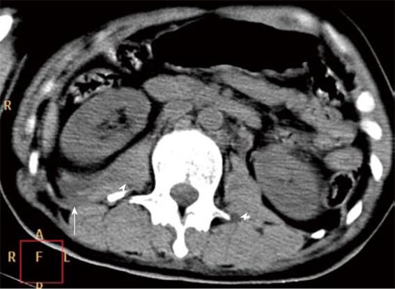
Lumbar vertebral crush fractures were detected in a total of 49 vertebrae in 47.06% (24/51) of patients. The fractures occurred in the transverse process (Figure 1), the body, spinous process or vertebral plate of lumber vertebrae. These crush fractures are listed in Table 1. Factures of the transverse process were more common than those of any other anatomic sites of a lumber vertebra (P < 0.001).
| Anatomic sites of vertebrae involved | Cases of lumbar vertebral fractures |
| Transverse process | 15 (62.5) |
| The body | 1 (4.17) |
| Transverse process + the body | 4 (16.67) |
| Transverse process + spinous process | 1 (4.17) |
| Transverse process + the body + spinous process | 1 (4.17) |
| Transverse process + the body + vertebral plate | 1 (4.17) |
In this cohort, the mean number of lumbar vertebrae involved per patient with lumbar vertebral crush fractures was 2 vertebrae with the peak prevalence between 1 and 2 of the number affected which ranged from 1 to 5. The cohort was composed of 33.33% of patients (8/24) with fractures of single vertebra and 66.67% (16/24) with fractures of multiple vertebrae. Multiple fractures occurred in two vertebrae in 62.5% of patients (10/16) and in more than two vertebrae in 37.5% (6/16) of patients.
According to the lumbar levels involved, the fractures occurred in L1 vertebra in 26.53% (13/49) of vertebrae, in L2 vertebra in 34.69% (17/49), in L3 vertebra in 20.41% (10/49), in L4 vertebra in 12.24% (6/49), and in L5 vertebra in 6.12% (3/49).
With regard to injuries of abdominal wall soft tissue, these occurred in a total of 28 patients. Intramuscular hematoma, muscular swelling or fatty edema were detected in 64.29% of patients (18/28), and 35.71% of patients (10/28) had both subcutaneous air collection and swelling of abdominal wall soft tissue. According to the anatomic regions involved, injuries of abdominal wall soft tissue occurred in the anterior abdominal wall in 10.71% of patients (3/28), in the posterior abdominal wall in 14.29% (4/28), in the flank abdominal wall in 24.43% (6/28), in both the anterior and posterior abdominal wall in 14.29% (4/28), in the flank and anterior abdominal wall in 10.71% (3/28), in the flank and posterior abdominal wall in 10.71% (3/28), and in the flank and anterior and posterior abdominal wall in 17.86% (5/28).
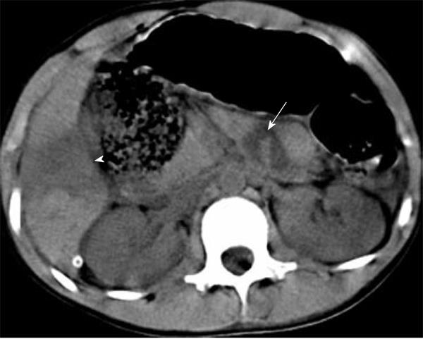
In this cohort, 29 patients had retroperitoneal injuries. The injuries were composed of renal or perirenal injuries (Figure 1), traumatic pancreatitis or pancreatic injury (Figure 2), rupture of abdominal aortic aneurysm, fluid collection in perirenal space and air collection in the posterior perirenal space in a total of 14 patients, and the remaining patients had swelling of perirenal fascia. The detailed results are listed in Table 2. Renal or perirenal injuries were more often seen than injuries of the other organs in the retroperitoneal space (P < 0.001).
| Retroperitoneal injuries | Cases of crush retroperitoneal injuries |
| Renal and perirenal injuries | |
| Perirenal hematoma | 4 (28.57) |
| Subcapsular hematoma | 1 (7.14) |
| Fluid collection in perirenal space | 2 (14.29) |
| Air collection in perirenal space | 1 (7.14) |
| Hemorrhage of renal cyst | 1 (7.14) |
| Pancreatic injury | |
| Traumatic pancreatitis | 2 (14.29) |
| Pancreatic rupture with hematoma | 1 (7.14) |
| Renal injuries + pancreatic injury | |
| Renal contusion + pancreatic contusion | 1 (7.14) |
| Renal injuries + injury of abdominal aort | |
| Rupture of abdominal aortic aneurysm + perirenal hematoma | 1 (7.14) |
As for intraperitoneal injuries, 16 patients had intraperitoneal injuries. Intraperitoneal parenchymal injuries were composed of liver injuries (Figure 2) and splenic injuries with or without seroperitoneum and pneumoperitoneum in a total of 9 patients, and the remaining patients were composed of seroperitoneum in 5 patients and pneumoperitoneum in 2 patients. Detailed cases of these results are listed in Table 3. Liver injuries were more often seen than splenic injuries (P < 0.001).
| Intraperitoneal injuries | Cases of crush intraperitoneal injuries |
| Liver injuries | |
| Hepatorrhexis | 3 (33.33) |
| Hepatorrhexis with hematoma | 3 (33.33) |
| Hepatorrhexis with subcapsular hematoma | 1 (11.11) |
| Splenic rupture | |
| With hematoma | 1 (11.11) |
| With subcapsular hematoma | 1 (11.11) |
Additionally, parenchymal polytraumatism (Figure 2) associated with the earthquake was found in 7.84% (4/51) of patients including injury in bilateral kidneys, in unilateral kidney and liver, in liver and pancreas, and in unilateral kidney and liver and pancreas each in 1 patient.
During the past 20 years, natural disasters have claimed more than three million lives worldwide[13]. With respect to loss of life, an earthquake is the most harmful disaster among all types of natural disasters[14]. In the 2008 Sichuan earthquake, 374 643 people were injured. Based on the patients presenting in the university hospital, abdominal crush injuries were one type of injury which occurred in 2.05% (56 of 2728), which was higher than the morbidity (1%) in the 2005 Kashmir earthquake[15]. Based on the largest number of injured people treated in the hospital in the earthquake-affected area, we proposed that the CT features of abdominal crush injuries might be helpful in better understanding and treatment planning of survivors with abdominal crush injuries in similar future earthquakes.
As an emergency imaging tool for diagnosing abdominal crush injuries, MDCT helps to image the injuries as soon as possible in a great number of injured patients due to an increased speed of image acquisition, and significantly reduces the time requirements for initial diagnostic evaluation[16]. We, therefore, focused on the emergency assessment of abdominal injuries using 16-row MDCT scanners in this earthquake setting. Because of the suspicion of acute renal failure associated with crush syndrome in the massive earthquake, use of iodinated contrast might presumably potentiate the development of renal failure in some patients, and the emergency CT scans of the abdomen were performed without intravenous contrast material.
Clinically, we found that fractures of lumbar vertebrae and injury of abdominal wall soft tissue were more often seen than retro- and intraperitoneal injuries, which may be explained by the fact that some victims with retro- and intraperitoneal injuries may have died before a rescue could be carried out, whereas most victims with fractures of lumbar vertebrae or injury of abdominal wall soft tissue survived long enough to be transported to the hospital to receive effective treatments.
Additionally, we found that retroperitoneal injuries were more common than intraperitoneal injuries. According to Chen et al[17], most victims fell down and were trapped in the prone position when the earthquake occurred, and the high-force impacts of collapsed buildings and falling objects frequently struck the lower back, which eventually results in a high frequency of retroperitoneal injuries.
With regard to fractures of lumbar vertebrae, fractures of multiple vertebrae were more often seen than fractures of single vertebra, and occurred most frequently in two vertebrae. Regarding the involved anatomic sites of a vertebra, fractures of the transverse process were more common than those of the body, spinous process and vertebral plate. Based on the vertebral levels involved, L1-3 fractures were more common in patients with crush fractures of lumbar vertebrae, which was consistent with a previous report[9].
As for retroperitoneal parenchymal injuries in the massive earthquake, renal and perirenal injuries were more often seen than injuries to other organs in the retroperitoneal space. According to Sever et al[18], the renal victims were of vital importance since they could predict the final outcome as well as be directive for medical therapies. We presumed that the CT findings could help us to better understand renal injuries from the earthquake for effective treatments to improve therapeutic outcome.
As for intraperitoneal parenchymal injuries arising from this earthquake, they were composed of liver and splenic injuries. Liver injuries were more often seen than injuries of any other parenchymal viscera. In addition, seroperitoneum occurred frequently in patients with abdominal crush injury. Full thickness bowel disruption was not represented in the population under study, which may be explained by the fact that victims with lethal injuries died before a rescue was carried out.
In view of the severity of abdominal crush injuries, abdominal parenchymal polytraumatism may be a good indicator. According to Ersoy et al[19], multiple injuries were more frequent in nonsurvivors than in survivors during the Marmara earthquake, and polytraumatism was one of the main factors which increased mortality risk in dialyzed injuries after the earthquake. In our study, we found that 7.84% (4/51) of patients had parenchymal polytraumatism. Although the morbidity of victims with abdominal parenchymal polytraumatism was not significant, attention should be paid clinically to prevent high mortality.
There were two inevitable limitations in our study. Firstly, contrast-enhanced abdominal CT scanning could not be performed in the earthquake situation due to suspicion of acute renal failure associated with crush syndrome, however, these non-enhanced CT scans could still image the injury patterns as illustrated in the present study. Secondly, this university hospital was 92 kilometers away from the epicenter, which could cause selection bias in the sampling of patients. Despite the limitations, this study illustrates the features of abdominal crush injury in survivors using MDCT focusing on the predominant anatomic distributions, which may be helpful in better understanding abdominal crush injury resulting from another earthquake to provide effective treatments.
In conclusion, the high incidence of lumbar vertebral fractures occurring predominantly in the transverse process of two vertebrae among L1-3 vertebrae, and in injuries of abdominal wall soft tissue; and a relatively high incidence of retroperitoneal parenchymal injury predominantly in kidney compared to intraperitoneal parenchymal injury predominantly in liver, might be features of abdominal crush injuries in an earthquake. We hope that these features of abdominal crush injuries may be helpful for the treatment of survivors in similar future earthquakes.
In the 2008 Sichuan earthquake, some patients sustained abdominal trauma associated with crush injury. Computed tomography (CT) scanning of the abdomen is currently considered the gold standard for detecting intra- and retroperitoneal lesions in trauma patients. However, the CT features of crush abdominal trauma in an earthquake have not been reported in the literature in detail.
Abdominal crush injuries resulting from the 2008 Sichuan earthquake shown on emergency non-enhanced multidetector CT (MDCT) images were reviewed focusing on the anatomic regions which included the lumbar vertebrae, abdominal wall soft tissue, retroperitoneum and intraperitoneal space; and the types of traumatic lesions.
A high incidence of abdominal wall soft tissue injuries and in fractures of lumbar vertebrae predominantly in the transverse process of two vertebrae among L1-3 vertebrae, and a relatively high incidence of retroperitoneal parenchymal injury predominantly in kidney compared to intraperitoneal parenchymal injury predominantly in liver may be features of abdominal crush injuries in an earthquake.
The features of abdominal crush injuries in the 2008 Sichuan earthquake would be helpful for better understanding and treatment planning of abdominal crush trauma survivors in similar future earthquakes.
Emergency non-enhanced MDCT is a rapid and valuable procedure to demonstrate abdominal crush injuries in an earthquake in detail, and is helpful for clinicians to better understand the features of abdominal crush injuries.
In this manuscript, the authors described the features of abdominal crush injuries in an earthquake, which is interesting.
Peer reviewer: Yasunori Minami, MD, PhD, Assistant Professor, Division of Gastroenterology and Hepatology, Department of Internal Medicine, Kinki University School of Medicine, 377-2 Ohno-higashi, Osaka-sayama, Osaka, 589-8511, Japan
S- Editor Cheng JX L- Editor Webster JR E- Editor Zheng XM
| 1. | Muckart DJ, Thomson SR. Undetected injuries: a preventable cause of increased morbidity and mortality. Am J Surg. 1991;162:457-460. [PubMed] [Cited in This Article: ] |
| 2. | Hamilton JD, Kumaravel M, Censullo ML, Cohen AM, Kievlan DS, West OC. Multidetector CT evaluation of active extravasation in blunt abdominal and pelvic trauma patients. Radiographics. 2008;28:1603-1616. [PubMed] [Cited in This Article: ] |
| 3. | Scaglione M. The use of sonography versus computed tomography in the triage of blunt abdominal trauma: the European perspective. Emerg Radiol. 2004;10:296-298. [PubMed] [Cited in This Article: ] |
| 4. | Yoshii H, Sato M, Yamamoto S, Motegi M, Okusawa S, Kitano M, Nagashima A, Doi M, Takuma K, Kato K. Usefulness and limitations of ultrasonography in the initial evaluation of blunt abdominal trauma. J Trauma. 1998;45:45-50; discussion 50-1. [PubMed] [Cited in This Article: ] |
| 5. | Federle MP. Computed tomography of blunt abdominal trauma. Radiol Clin North Am. 1983;21:461-475. [PubMed] [Cited in This Article: ] |
| 6. | Federle MP, Griffiths B, Minagi H, Jeffrey RB. Splenic trauma: evaluation with CT. Radiology. 1987;162:69-71. [PubMed] [Cited in This Article: ] |
| 7. | Becker CD, Mentha G, Terrier F. Blunt abdominal trauma in adults: role of CT in the diagnosis and management of visceral injuries. Part 1: liver and spleen. Eur Radiol. 1998;8:553-562. [PubMed] [Cited in This Article: ] |
| 8. | Mirvis SE, Shanmuganathan K. Trauma radiology: part I. Computerized tomographic imaging of abdominal trauma. J Intensive Care Med. 1994;9:151-163. [PubMed] [Cited in This Article: ] |
| 9. | Dong ZH, Yang ZG, Chen TW, Feng YC, Wang QL, Chu ZG. Spinal injuries in the Sichuan earthquake. N Engl J Med. 2009;361:636-637. [PubMed] [Cited in This Article: ] |
| 10. | Dong ZH, Yang ZG, Chen TW, Feng YC, Chu ZG, Yu JQ, Bai HL, Wang QL. Crush thoracic trauma in the massive Sichuan earthquake: evaluation with multidetector CT of 215 cases. Radiology. 2010;254:285-291. [PubMed] [Cited in This Article: ] |
| 11. | Chen TW, Yang ZG, Dong ZH, Chu ZG, Yao J, Wang QL. Pelvic crush fractures in survivors of the Sichuan earthquake evaluated by digital radiography and multidetector computed tomography. Skeletal Radiol. 2010;39:1117-1122. [PubMed] [Cited in This Article: ] |
| 12. | Chen TW, Yang ZG, Wang QL, Dong ZH, Yu JQ, Zhuang ZP, Hou CL, Li ZL. Crush extremity fractures associated with the 2008 Sichuan earthquake: anatomic sites, numbers and statuses evaluated with digital radiography and multidetector computed tomography. Skeletal Radiol. 2009;38:1089-1097. [PubMed] [Cited in This Article: ] |
| 13. | Peek-Asa C, Kraus JF, Bourque LB, Vimalachandra D, Yu J, Abrams J. Fatal and hospitalized injuries resulting from the 1994 Northridge earthquake. Int J Epidemiol. 1998;27:459-465. [PubMed] [Cited in This Article: ] |
| 14. | Mahoney LE, Reutershan TP. Catastrophic disasters and the design of disaster medical care systems. Ann Emerg Med. 1987;16:1085-1091. [PubMed] [Cited in This Article: ] |
| 15. | Mulvey JM, Awan SU, Qadri AA, Maqsood MA. Profile of injuries arising from the 2005 Kashmir earthquake: the first 72 h. Injury. 2008;39:554-560. [PubMed] [Cited in This Article: ] |
| 16. | Hessmann MH, Hofmann A, Kreitner KF, Lott C, Rommens PM. The benefit of multislice CT in the emergency room management of polytraumatized patients. Acta Chir Belg. 2006;106:500-507. [PubMed] [Cited in This Article: ] |
| 17. | Chen TW, Yang ZG, Dong ZH, Chu ZG, Tang SS, Deng W. Earthquake-related crush injury versus non-earthquake injury in abdominal trauma patients on emergency multidetector computed tomography: a comparative study. J Korean Med Sci. 2011;26:438-443. [PubMed] [Cited in This Article: ] |
| 18. | Sever MS, Erek E, Vanholder R, Akoglu E, Yavuz M, Ergin H, Turkmen F, Korular D, Yenicesu M, Erbilgin D. Clinical findings in the renal victims of a catastrophic disaster: the Marmara earthquake. Nephrol Dial Transplant. 2002;17:1942-1949. [PubMed] [Cited in This Article: ] |
| 19. | Ersoy A, Yavuz M, Usta M, Ercan I, Aslanhan I, Güllülü M, Kurt E, Emir G, Dilek K, Yurtkuran M. Survival analysis of the factors affecting in mortality in injured patients requiring dialysis due to acute renal failure during the Marmara earthquake: survivors vs non-survivors. Clin Nephrol. 2003;59:334-340. [PubMed] [Cited in This Article: ] |


