Autobiography of Editorial Board Members Open Access
Copyright ©2011 Baishideng Publishing Group Co., Limited. All rights reserved.
World J Radiol. Apr 28, 2011; 3(4): 120-124
Published online Apr 28, 2011. doi: 10.4329/wjr.v3.i4.120
Feng Chen’s work on translational and clinical imaging
Feng Chen, Section of Radiology, Department of Medical Diagnostic Sciences, Faculty of Medicine, University of Leuven, 3000 Leuven, Belgium
Feng Chen, Molecular Small Animal Imaging Center, University of Leuven, 3000 Leuven, Belgium
Feng Chen, Department of Radiology, Zhong Da Hospital, Southeast University, Nanjing 210009, Jiangsu Province, China
Author contributions: Chen F solely contributed to this manuscript.
Supported by Health Bureau of Jiangsu Province, China; Chinese Scholarship Council; National Natural Science Foundation of China; European Congress of Radiology 2000, EAR-ECR Research and Education Fund Fellowship Grant; European Union Asia-Link Project
Correspondence to: Feng Chen, MD, PhD, Section of Radiology, Department of Medical Diagnostic Sciences, Faculty of Medicine, University of Leuven, Herestraat 49, bus 7003, 3000 Leuven, Belgium. chenfengbe@yahoo.com.cn
Telephone: +32-16-330165 Fax: +32-16-343765
Received: October 11, 2010
Revised: March 28, 2011
Accepted: April 4, 2011
Published online: April 28, 2011

Abstract

Dr. Feng Chen is a chief medical doctor and the vice chairman of the Department of Radiology in Zhong Da Hospital at Southeast University, Nanjing, China and a senior researcher in the Department of Radiology at the Catholic University of Leuven, Belgium. His main areas of interest are translational imaging research including stroke, tumor angiogenesis, assessment of therapeutic response in solid tumors, and magnetic resonance contrast media. Dr. Feng Chen has published 44 scientific papers in peer-reviewed international journals. He and his colleagues have developed an imaging platform which includes animal models, animal preparations and multiparametric magnetic resonance imaging (MRI) protocols for translational animal imaging research using clinical machines. His MRI findings on rodent stroke are considered to “serve as a model for future laboratory investigations of treatment of acute stroke and unify the approaches developed for clinical studies”. He and his colleagues have introduced a novel liver tumor model in rodents, in which a series of studies concerning the antitumor activity of vascular disrupting agents have been successively conducted and assessed by in vivo MRI, especially by diffusion weighted imaging as an imaging biomarker. His goal is to provide valuable references for clinical practice and to contribute to the translation of animal imaging research into patient applications.

Key Words: Animal study, Contrast agent, Magnetic resonance imaging, Therapeutic assessment, Translational research, Tumor angiogenesis, Tumor therapy, Vascular disrupting agent


INTRODUCTION AND EDUCATIONAL EXPERIENCE

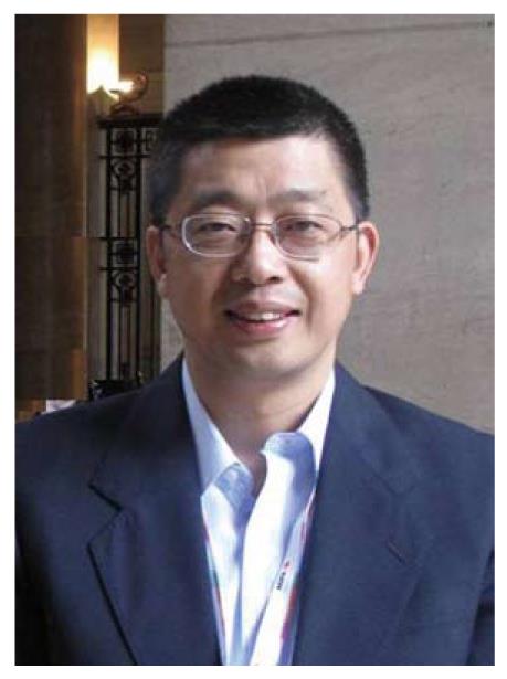
Dr. Feng Chen is a chief medical doctor and the vice chairman of the Department of Radiology in Zhong Da Hospital at Southeast University, Nanjing, China. Currently, he is also a senior researcher in the Department of Radiology at the Catholic University of Leuven, Belgium (Figure 1). He obtained his Bachelor’s degree in Medicine in 1984 and Master’s degree in Radiology in 1987 from Nanjing Railway Medical College (known as Medical School of Southeast University since 2000), China. He was employed as a radiologist in Zhong Da Hospital of Southeast University from 1987 to 2001. He was awarded a Chinese Overseas Scholarship to support his clinical magnetic resonance imaging (MRI) research at Leeds University in the UK as a visiting scholar from 1997 to 1998. He obtained his PhD degree from the Catholic University of Leuven in 2007. Since then he has been employed at the University of Leuven.

Figure 1
Figure 1 Feng Chen, MD, PhD, Section of Radiology, Department of Medical Diagnostic Sciences, Faculty of Medicine, University of Leuven, Herestraat 49, bus 7003, 3000 Leuven, Belgium.

Dr. Feng Chen has published 44 scientific papers in peer-reviewed international journals. He has accepted several invitations to be a panel expert at international cancer conferences, to speak at international meetings, and to write reviews or chapters on small animal imaging and oncology imaging using clinical MR scanners. Throughout his academic career, he has been supported by a series of awards including the Chinese Scholarship Council; National Natural Science Foundation of China; EAR-ECR Research and Education Fund Fellowship Grant, European Congress of Radiology 2000, and European Union Asia-Link Program. He was also the silver prize winner of the QUIZ on Asian-Oceanian Seminars on Abdominal Radiology in 1997 and the silver prize winner of the QUIZ on Diagnostic and Interventional Radiology in 1999.

ACADEMIC STRATEGY AND GOALS

As a radiologist with more than 15 years work experience, Dr. Chen is well aware of the vital role of preclinical imaging research in studying the mechanism of human pathologies and in finding a solution for problems encountered in daily clinical practice. Therefore, over the last 9 years Dr. Chen’s main work has been focused on translational imaging research[1-15]. Since clinical MR scanners are available to most institutions conducting animal experiments, he and his colleagues have developed a platform which includes animal models, animal preparations and a multiparametric MRI protocol for translational animal imaging research using clinical machines[16]. Based on this imaging platform, a number of animal models such as the modified photothrombotic stroke model[1], implanted liver tumor model[4,17] and reperfused partial liver infarction model in rodents[11] have been characterized, and the therapeutic efficacy of some novel thrombolytic and anticancer agents have been monitored and evaluated[9,12,18]. Dr. Chen is also pursuing the further investigation of ischemic diseases and tumor angiogenesis imaging, and exploring novel specific imaging and biological markers for the noninvasive assessment of pathological processes. The goal is to provide valuable references for clinical practice and to contribute to the translation of animal imaging research into patient applications.

ACADEMIC ACHIEVEMENTS
MRI studies in rodent stroke

During the 5-year period of his PhD program, Dr. Chen systematically studied a model of photochemically-induced thrombosis (PIT) of proximal middle cerebral artery occlusion with MRI using a 1.5 Tesla clinical scanner[1-3,7,8,19]. With this model, the therapeutic effects of a novel anti-stroke agent, microplasmin (μPli) has been monitored and evaluated with MRI, and compared with the approved thrombolytic agent, tissue plasminogen activator (tPA)[9]. The results indicated that μPli with superior safety may be a potential alternative to tPA for the treatment of focal ischemic stroke.

The main findings of this series of studies included: (1) An ischemic penumbra defined with diffusion and perfusion mismatch using MRI has been approved in this PIT stroke model, which is used as an inclusion criterion before treatment[6,9]; (2) A delayed perfusion phenomenon has been observed for the first time in a stroke animal model by MR perfusion imaging, which plays an important role in maintaining perfusion to the “penumbra” region after stroke onset[3,8]; and (3) The evidence of μPli in the treatment of the stroke model has been documented with noninvasive multiparametric MRI[9,20].

The findings of Dr. Chen and his colleagues “demonstrate the ability to identify the ischemic and putatively infarcted regions before therapy, to institute therapy in a model that compares thrombolytic agents, and to document the response and complication rates. These findings serve as a model for future laboratory investigations of the treatment of acute stroke and unify the approaches developed for clinical studies” as stated in an Editorial on Dr. Chen and his colleague’s article which appeared in an issue of Radiology[21].

Imaging assessment of therapeutic effects in oncology

Dr. Chen’s research activities and contribution in this field are mainly associated with the following work:

Novel liver implanted tumor models in rodents were developed and characterized using comprehensive MRI techniques which included morphologic, functional, and metabolic information[4,17]. These models provided an upgraded research platform for the preclinical assessment of diagnostic and therapeutic strategies as indicated by the consequent outcomes listed below.

Diffusion weighted imaging (DWI) has emerged as a unique and powerful non-invasive MRI technique. Its value as an imaging biomarker has been investigated extensively in imaging-based diagnosis in a variety of translational applications including cerebral ischemia and solid tumors using small animals. Through technical adaptation and optimization, Dr. Chen and his colleagues have been able to perform clinically relevant animal studies with conclusions based on DWI quantification using a clinical magnet[5,22].

MRI monitoring and evaluation of experimental hepatic rhabdomyosarcoma treated with a vascular disrupting agent (VDA) has been conducted in a series of studies[12-15,18]. The results demonstrate that clinical MRI allows monitoring of VDA-related vascular shutdown and the discrimination between viable tissue and necrotic tumor tissue. However, a single dose of VDA seems insufficient for tumor eradication due to evident peripheral residue and recurrence[23]. Therefore, VDA combined with other therapeutic strategies, such as antiangiogenics, have been proposed and are undergoing investigation, and are believed to be more easily translated into patient practice because of the clinically relevant experimental set-up.

Investigation and application of tissue specific MR contrast agents

Necrosis-avid contrast agents (NACAs) have thus far been discovered and developed by Prof. Ni in the Catholic University of Leuven, Belgium as necrosis-targeting markers for MRI identification of non-viable tissue[24]. During recent years, Dr. Chen has been actively involved in the investigation of NACAs[25,26] and their application in acute myocardial infarction[27], tissue viability assessment, and therapeutic evaluation after interventional therapies[28].

Dr. Chen also compared the effects of two superparamagnetic iron oxide (SPIO) contrast agents, ferumoxides and SHU-555A, in MRI of liver and spleen in the early phase of their clinical application[29], which provided valuable information for best practice in patients.

Computed tomography gastrography for diagnosis of gastric carcinoma

Gastric carcinoma is one of the leading causes of death in East Asia. Dr Chen was awarded an EAR-ECR Fellowship Grant in this field by the Research and Education Fund during the European Congress of Radiology 2000 in Vienna. In this study, two- and three-dimensional display techniques after spiral computed tomography (CT) scanning were cross-referenced. The role of the combined CT technique was compared with that of upper gastrointestinal series, fiberoptic gastroscopy and histopathology in the detection, Borrmann’s classification, and staging of gastric carcinoma[30]. Based on the work described above, Dr. Chen, together with two colleagues, edited a book entitled “Virtual Endoscopy and Related 3D in Clinical Medicine”[31]. Dr. Chen also systematically introduced the diagnostic test, the receiver operative characteristic curve (ROC)[32] approach in diagnostic radiology in China.

Imaging study on relationship between anomalous junction of pancreaticobiliary duct and diseases of biliary tract and pancreas

Dr. Chen systematically investigated and reported, for the first time, the occurrence of anomalous junction of the pancreaticobiliary duct (AJPBD) based on cholangiopancreatography in a Chinese population. The results showed that AJPBD is a possible risk factor for biliary duct and pancreatic diseases. A new complex pattern of AJPBD was proposed and a novel technique for magnetic resonance pancreatography after oral juice stimulation was developed. Dr. Chen’s study findings have been listed as one of the most important advances in abdominal radiology in China[33].

PERSPECTIVE

Medical imaging is a rapidly growing field in medicine. Small animal imaging has becoming a major player in an increasing number of animal experiments in which MRI has been a favorite choice for in vivo monitoring due to its advantages which include excellent resolution and innocuousness[34]. As one of his research interests, Dr. Chen’s work will focus on the novel anti-tumor strategies involving the application of VDA assessed by in vivo imaging techniques. Antivascular tumor therapies with VDAs are a promising approach in oncologic research and have been conducted in a number of studies[5,12,14,18]. However, rapid tumor re-growth from a residual viable rim after treatment with a VDA compromises the therapeutic efficacy of these agents[23]. Several approaches have been suggested to solve this problem including biological and targeted radiotherapy methods. For instance, tumor rebound after VDA treatment is reported to be associated with the acute mobilization of bone marrow (BM)-derived circulating endothelial progenitor cells (EPCs) induced by VDA, which contributes to the responsive tumor neoangiogenesis. The combined use of antiangiogenics with VDA may inhibit the mobilization of EPCs so that tumor recurrence may be prevented or reduced[35]. However, critical controversy exists in the literature regarding this theory and the consequent role of EPCs in tumor growth[36]. Therefore, exploration of this scientific issue may allow better insight into the mechanism of tumor recurrence, and lead to new anti-tumor strategies and promote imaging research.

Footnotes

Peer reviewer: Roberto Grassi, MD, Full Professor of Radiology, Department “ Magrassi-Lanzara ” Institute of Radiology, Second University of Naples, Piazza Miraglia, Naples, 80138, Italy

S- Editor Cheng JX L- Editor Webster JR E- Editor Zheng XM

References
1.  Chen F, Suzuki Y, Nagai N, Peeters R, Coenegrachts K, Coudyzer W, Marchal G, Ni Y. Visualization of stroke with clinical MR imagers in rats: a feasibility study. Radiology. 2004;233:905-911.  [PubMed]  [DOI]  [Cited in This Article: ]
2.  Chen F, Suzuki Y, Nagai N, Peeters R, Sun X, Coudyzer W, Marchal G, Ni Y. Rat cerebral ischemia induced with photochemical occlusion of proximal middle cerebral artery: a stroke model for MR imaging research. MAGMA. 2004;17:103-108.  [PubMed]  [DOI]  [Cited in This Article: ]
3.  Chen F, Suzuki Y, Nagai N, Peeters R, Marchal G, Ni Y. Dynamic susceptibility contrast-enhanced perfusion MR imaging at 1.5 T predicts final infarct size in a rat stroke model. J Neurosci Methods. 2005;141:55-60.  [PubMed]  [DOI]  [Cited in This Article: ]
4.  Chen F, Sun X, De Keyzer F, Yu J, Peeters R, Coudyzer W, Vandecaveye V, Landuyt W, Bosmans H, Van Hecke P. Liver tumor model with implanted rhabdomyosarcoma in rats: MR imaging, microangiography, and histopathologic analysis. Radiology. 2006;239:554-562.  [PubMed]  [DOI]  [Cited in This Article: ]
5.  Chen F, De Keyzer F, Wang H, Vandecaveye V, Landuyt W, Bosmans H, Hermans R, Marchal G, Ni Y. Diffusion weighted imaging in small rodents using clinical MRI scanners. Methods. 2007;43:12-20.  [PubMed]  [DOI]  [Cited in This Article: ]
6.  Chen F, Liu Q, Wang H, Suzuki Y, Nagai N, Yu J, Marchal G, Ni Y. Comparing two methods for assessment of perfusion-diffusion mismatch in a rodent model of ischaemic stroke: a pilot study. Br J Radiol. 2008;81:192-198.  [PubMed]  [DOI]  [Cited in This Article: ]
7.  Chen F, Suzuki Y, Nagai N, Jin L, Yu J, Wang H, Marchal G, Ni Y. Rodent stroke induced by photochemical occlusion of proximal middle cerebral artery: evolution monitored with MR imaging and histopathology. Eur J Radiol. 2007;63:68-75.  [PubMed]  [DOI]  [Cited in This Article: ]
8.  Chen F, Suzuki Y, Nagai N, Sun X, Coudyzer W, Yu J, Marchal G, Ni Y. Delayed perfusion phenomenon in a rat stroke model at 1.5 T MR: an imaging sign parallel to spontaneous reperfusion and ischemic penumbra? Eur J Radiol. 2007;61:70-78.  [PubMed]  [DOI]  [Cited in This Article: ]
9.  Chen F, Suzuki Y, Nagai N, Sun X, Wang H, Yu J, Marchal G, Ni Y. Microplasmin and tissue plasminogen activator: comparison of therapeutic effects in rat stroke model at multiparametric MR imaging. Radiology. 2007;244:429-438.  [PubMed]  [DOI]  [Cited in This Article: ]
10.  De Keyzer F, Vandecaveye V, Thoeny H, Chen F, Ni Y, Marchal G, Hermans R, Nuyts S, Landuyt W, Bosmans H. Dynamic contrast-enhanced and diffusion-weighted MRI for early detection of tumoral changes in single-dose and fractionated radiotherapy: evaluation in a rat rhabdomyosarcoma model. Eur Radiol. 2009;19:2663-2671.  [PubMed]  [DOI]  [Cited in This Article: ]
11.  Wu X, Wang H, Chen F, Jin L, Li J, Feng Y, DeKeyzer F, Yu J, Marchal G, Ni Y. Rat model of reperfused partial liver infarction: characterization with multiparametric magnetic resonance imaging, microangiography, and histomorphology. Acta Radiol. 2009;50:276-287.  [PubMed]  [DOI]  [Cited in This Article: ]
12.  Wang H, Sun X, Chen F, De Keyzer F, Yu J, Landuyt W, Vandecaveye V, Peeters R, Bosmans H, Hermans R. Treatment of rodent liver tumor with combretastatin a4 phosphate: noninvasive therapeutic evaluation using multiparametric magnetic resonance imaging in correlation with microangiography and histology. Invest Radiol. 2009;44:44-53.  [PubMed]  [DOI]  [Cited in This Article: ]
13.  Thoeny HC, De Keyzer F, Chen F, Ni Y, Landuyt W, Verbeken EK, Bosmans H, Marchal G, Hermans R. Diffusion-weighted MR imaging in monitoring the effect of a vascular targeting agent on rhabdomyosarcoma in rats. Radiology. 2005;234:756-764.  [PubMed]  [DOI]  [Cited in This Article: ]
14.  Thoeny HC, De Keyzer F, Vandecaveye V, Chen F, Sun X, Bosmans H, Hermans R, Verbeken EK, Boesch C, Marchal G. Effect of vascular targeting agent in rat tumor model: dynamic contrast-enhanced versus diffusion-weighted MR imaging. Radiology. 2005;237:492-499.  [PubMed]  [DOI]  [Cited in This Article: ]
15.  Thoeny HC, De Keyzer F, Chen F, Vandecaveye V, Verbeken EK, Ahmed B, Sun X, Ni Y, Bosmans H, Hermans R. Diffusion-weighted magnetic resonance imaging allows noninvasive in vivo monitoring of the effects of combretastatin a-4 phosphate after repeated administration. Neoplasia. 2005;7:779-787.  [PubMed]  [DOI]  [Cited in This Article: ]
16.  Chen F, De Keyzer F, Ni Y. Cancer Models - Multiparametric Applications of Clinical MRI in Rodent Hepatic Tumor Model. In vivo NMR imaging: methods and protocols. New York: Humana Press 2011; In press.  [PubMed]  [DOI]  [Cited in This Article: ]
17.  Wang H, Van de Putte M, Chen F, De Keyzer F, Jin L, Yu J, Marchal G, de Witte P, Ni Y. Murine liver implantation of radiation-induced fibrosarcoma: characterization with MR imaging, microangiography and histopathology. Eur Radiol. 2008;18:1422-1430.  [PubMed]  [DOI]  [Cited in This Article: ]
18.  Wang H, Li J, Chen F, De Keyzer F, Yu J, Feng Y, Nuyts J, Marchal G, Ni Y. Morphological, functional and metabolic imaging biomarkers: assessment of vascular-disrupting effect on rodent liver tumours. Eur Radiol. 2010;20:2013-2026.  [PubMed]  [DOI]  [Cited in This Article: ]
19.  Chen F, Marchal G, Ni Y. Rodent models and magnetic resonance imaging: diagnostic and therapeutic utilities for stroke. Int J Model Ident Contr. 2010;9:275-287.  [PubMed]  [DOI]  [Cited in This Article: ]
20.  Suzuki Y, Chen F, Ni Y, Marchal G, Collen D, Nagai N. Microplasmin reduces ischemic brain damage and improves neurological function in a rat stroke model monitored with MRI. Stroke. 2004;35:2402-2406.  [PubMed]  [DOI]  [Cited in This Article: ]
21.  Hackney DB. Does MR imaging improve precision in stroke thrombolysis trials? Radiology. 2007;244:323-324.  [PubMed]  [DOI]  [Cited in This Article: ]
22.  Sun X, Wang H, Chen F, De Keyzer F, Yu J, Jiang Y, Feng Y, Li J, Marchal G, Ni Y. Diffusion-weighted MRI of hepatic tumor in rats: comparison between in vivo and postmortem imaging acquisitions. J Magn Reson Imaging. 2009;29:621-628.  [PubMed]  [DOI]  [Cited in This Article: ]
23.  Tozer GM, Kanthou C, Baguley BC. Disrupting tumour blood vessels. Nat Rev Cancer. 2005;5:423-435.  [PubMed]  [DOI]  [Cited in This Article: ]
24.  Ni Y, Bormans G, Chen F, Verbruggen A, Marchal G. Necrosis avid contrast agents: functional similarity versus structural diversity. Invest Radiol. 2005;40:526-535.  [PubMed]  [DOI]  [Cited in This Article: ]
25.  Van de Putte M, Wang H, Chen F, de Witte PA, Ni Y. Hypericin as a marker for determination of tissue viability after intratumoral ethanol injection in a murine liver tumor model. Acad Radiol. 2008;15:107-113.  [PubMed]  [DOI]  [Cited in This Article: ]
26.  Ni Y, Huyghe D, Verbeke K, de Witte PA, Nuyts J, Mortelmans L, Chen F, Marchal G, Verbruggen AM, Bormans GM. First preclinical evaluation of mono-[123I]iodohypericin as a necrosis-avid tracer agent. Eur J Nucl Med Mol Imaging. 2006;33:595-601.  [PubMed]  [DOI]  [Cited in This Article: ]
27.  Jin J, Teng G, Feng Y, Wu Y, Jin Q, Wang Y, Wang Z, Lu Q, Jiang Y, Wang S. Magnetic resonance imaging of acute reperfused myocardial infarction: intraindividual comparison of ECIII-60 and Gd-DTPA in a swine model. Cardiovasc Intervent Radiol. 2007;30:248-256.  [PubMed]  [DOI]  [Cited in This Article: ]
28.  Ni Y, Chen F, Mulier S, Sun X, Yu J, Landuyt W, Marchal G, Verbruggen A. Magnetic resonance imaging after radiofrequency ablation in a rodent model of liver tumor: tissue characterization using a novel necrosis-avid contrast agent. Eur Radiol. 2006;16:1031-1040.  [PubMed]  [DOI]  [Cited in This Article: ]
29.  Chen F, Ward J, Robinson PJ. MR imaging of the liver and spleen: a comparison of the effects on signal intensity of two superparamagnetic iron oxide agents. Magn Reson Imaging. 1999;17:549-556.  [PubMed]  [DOI]  [Cited in This Article: ]
30.  Chen F, Ni YC, Zheng KE, Ju SH, Sun J, Ou XL, Xu MH, Zhang H, Marchal G. Spiral CT in gastric carcinoma: comparison with barium study, fiberoptic gastroscopy and histopathology. World J Gastroenterol. 2003;9:1404-1408.  [PubMed]  [DOI]  [Cited in This Article: ]
31.  Yang XJ, Chen F, Han P.  Virtual Endoscopy and Related 3D Imaging in Clinical Medicine. Beijing: People's Medical Publishing House 2000; .  [PubMed]  [DOI]  [Cited in This Article: ]
32.  Ward J, Chen F, Guthrie JA, Wilson D, Lodge JP, Wyatt JI, Robinson PJ. Hepatic lesion detection after superparamagnetic iron oxide enhancement: comparison of five T2-weighted sequences at 1.0 T by using alternative-free response receiver operating characteristic analysis. Radiology. 2000;214:159-166.  [PubMed]  [DOI]  [Cited in This Article: ]
33.  Xie J. Progress of abdominal radiology in recent 50 years in China. Zhonghua Fangshexue Zazhi. 1999;33:23-27.  [PubMed]  [DOI]  [Cited in This Article: ]
34.  Brockmann MA, Kemmling A, Groden C. Current issues and perspectives in small rodent magnetic resonance imaging using clinical MRI scanners. Methods. 2007;43:79-87.  [PubMed]  [DOI]  [Cited in This Article: ]
35.  Shaked Y, Ciarrocchi A, Franco M, Lee CR, Man S, Cheung AM, Hicklin DJ, Chaplin D, Foster FS, Benezra R. Therapy-induced acute recruitment of circulating endothelial progenitor cells to tumors. Science. 2006;313:1785-1787.  [PubMed]  [DOI]  [Cited in This Article: ]
36.  Purhonen S, Palm J, Rossi D, Kaskenpää N, Rajantie I, Ylä-Herttuala S, Alitalo K, Weissman IL, Salven P. Bone marrow-derived circulating endothelial precursors do not contribute to vascular endothelium and are not needed for tumor growth. Proc Natl Acad Sci USA. 2008;105:6620-6625.  [PubMed]  [DOI]  [Cited in This Article: ]