Published online Jun 28, 2025. doi: 10.4329/wjr.v17.i6.106582
Revised: April 18, 2025
Accepted: May 21, 2025
Published online: June 28, 2025
Processing time: 117 Days and 4.7 Hours
The article "Assessment of superior mesenteric vascular flow quantitation in children using four-dimensional flow magnetic resonance imaging" suggests to use of four-dimensional (4D) flow magnetic resonance imaging (MRI) which is also to measure the blood flow in the superior mesenteric vein (SMV) in pediatric patients over the traditional method. The study focuses on assessing the potential of SMV and superior mesenteric artery (SMA) flow quantification in children utilizing 4D flow MRI. It included 9 pediatric patients aged 18 years and below where 5 were male and 4 were female patients, on whom magnetic resonance enterorrhaphy (MRE) with 4D flow MRI protocol was used. Statistical analysis was performed using MedCalc. Measurements of SMV and SMA between two readers were calculated using Bland-Altman analysis. The results stated that six patients showed no MRE evidence of active inflammatory bowel disease, two patients showed unmarkable bowel appearance on MRI and one patient showed normal MRE without endoscopy performed at the same timeframe. The study utilized available 4D flow MRI sequences in this study aiming to show the fea
Core Tip: This study determines blood flow in children's abdomens using a unique form of magnetic resonance imaging (MRI) known as four-dimensional (4D) flow. The superior mesenteric artery and vein were the primary targets of the medical team's attention. This method does not involve radiation; therefore, it is safer than traditional X-rays. It also depicts the direction and speed of blood flow. There were nine youngsters who were examined, and the findings demonstrated that 4D flow MRI can assist in the early detection of stomach disorders. It has the potential to improve pediatric care while decreasing expenses and dangers. The 4D flow MRI could prove rather useful in treating intestinal problems with future investigation.
- Citation: Mukundan A, Gupta D, Karmakar R, Wang HC. Transforming pediatric imaging: The role of four-dimensional flow magnetic resonance imaging in quantifying mesenteric blood flow. World J Radiol 2025; 17(6): 106582
- URL: https://www.wjgnet.com/1949-8470/full/v17/i6/106582.htm
- DOI: https://dx.doi.org/10.4329/wjr.v17.i6.106582
Inflammatory bowel disease (IBD) and other complicated gastrointestinal illnesses in children provide considerable difficulties for healthcare providers and patients together[1]. It can be challenging to diagnose these conditions at an early stage, and they can be even more challenging to monitor over time, particularly in children who may not tolerate certain procedures or extended hospital visits. Measurements of blood flow in the intestines supply and drain arteries, superior mesenteric artery (SMA) and veins, are crucial to the correct diagnosis of bowel diseases[2]. Doppler ultrasonography, computed tomography (CT), and traditional magnetic resonance imaging (MRI) are common examples of traditional imaging methods utilized to evaluate this vascular flow. Doppler ultrasonography[3], for example, does not use ionizing radiation, is portable, and is easily accessible. However, it may not work as well on kids with bigger bodies or who can't stay still during the process, which can cause motion artifacts and make it harder to see deeper vessels. CT scans, on the other hand, show a lot more about anatomy but expose people to radiation, which is especially dangerous for kids because they are more sensitive and have longer life span. Although conventional MRI may provide radiation-free high-resolution pictures, the information on time-resolved blood flow that is essential for accurate vascular evaluation is inconsistently captured by conventional two-dimensional or three-dimensional (3D) methods.
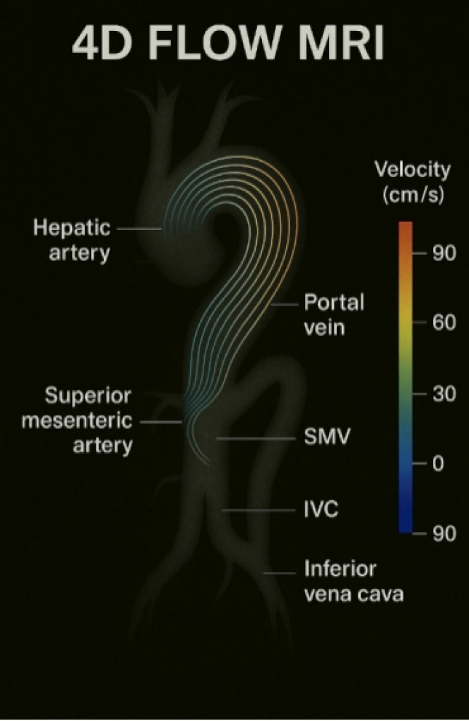
Four-dimensional (4D) flow MRI[4,5] has developed as a fresh method against this background. This sophisticated technique records 3D photographs of blood flow over time, therefore producing a "dynamic" dataset displaying not only the structure but also the direction and speed of blood inside specified veins. Figure 1 illustrates a representative 4D flow MRI visualization of mesenteric vasculature, including color-coded velocity mapping in key abdominal vessels. Researchers in a recent feasibility study sought to investigate if 4D flow MRI could be utilized to get reliable, repeatable measures of blood flow in the SMA and superior mesenteric vein (SMV)[6] of children patients either having bowel illnesses or suspected of having them. The 4D flow MRI[7] is a safer long-term choice for repeated tests as it uses no radiation unlike conventional techniques. It also provides thorough information on flow patterns that would enable the identification of minor anomalies associated with gut inflammation or vascular impairment.
Using this approach, the scientists retroactively examined nine children—between the ages of seven and fourteen, all of whom had magnetic resonance enterorrhaphy[8] combined with a 4D flow technique. For both the SMA and SMV, the researchers could record velocity and peak speed values at many segments—proximal, mid, and distal—by including 4D flow MRI into the usual imaging session. They then computed the time-resolved flow characteristics by centering an area of interest at each assigned segment. Independent evaluation of the pictures by two highly experienced radiologists using statistical techniques including Bland-Altman analysis[9] helped to ascertain the degree of agreement between these readers. The credibility of the method depends on this direct comparison. Clinical value is reduced if measurements differ much depending on the picture interpretation by someone. The study revealed strong interobserver agreement, however, which suggests that 4D flow MRI can in fact generate consistent and repeatable findings. Not just for its safety profile but also for the rich quantitative data it offers, 4D flow MRI stands out among conventional imaging modalities. Although Doppler ultrasounds can provide a moment of velocity at a given angle, the whole vessel and branching network are not as clearly shown. Although CT angiography[10] provides high anatomical information, every scan contains ionizing radiation and numerous phases may be required to show arterial and venous flow, hence increasing radiation exposure. Although standard MRI may record anatomy and some functional information, frequently it takes many distinct sequences to piece together what 4D flow MRI can achieve in one integrated protocol. Thus, this new technique might become a more effective and potent tool for assessing vascular flow in young children, thereby reducing both radiation and repetitive sedation or scanning sessions. The findings of the study also shed light on normal and maybe aberrant flow patterns. The scientists noted, for example, that whilst the SMA's velocity dropped, SMV flow velocity usually rose from the proximal to the distal segments. One day, this result may be used as a reference to spot aberrant variations in juvenile bowel illnesses. Furthermore, correlations with endoscopic evaluations showed that, when taken in line with clinical data, 4D flow MRI[11] measurements could help differentiate active disease from quiescent conditions; hence, more research with larger cohorts will be required to validate any diagnostic threshold. Beyond gastrointestinal applications, 4D flow MRI has shown clinical promise in several other pediatric settings. For instance, in congenital heart disease, it enables dynamic visualization of intracardiac and great vessel flow patterns, which assists in surgical planning and postoperative monitoring without the need for invasive catheterization or radiation exposure. Similarly, in pediatric neurovascular imaging, 4D flow MRI has been applied to assess cerebral venous outflow and intracranial hemodynamics in conditions such as venous sinus thrombosis or hydrocephalus. These applications demonstrate a broader trend of this modality transitioning from advanced research into routine diagnostic workflows across diverse pediatric specialties, supporting its integration into comprehensive child-centered imaging protocols. Beyond general assessments of bowel perfusion, 4D flow MRI holds significant promise in several specific pediatric gastrointestinal conditions. In IBD, quan
All things considered, 4D flow MRI presents a bright new path for assessing mesenteric arteries in young people. Its capacity to offer precise flow measurement free of radiation fits very nicely with the changing focus in pediatric medicine on patient safety and tailored treatment. These results pave the path for more general usage of 4D flow MRI even if the sample size was small and more study is required to completely establish its clinical benefit. This approach should greatly increase diagnostic accuracy, guide therapy decisions, and lower the requirement for invasive or repetitive imaging tests in juvenile bowel illness by allowing thorough hemodynamic evaluations in young patients with low risk.
The 4D flow MRI presents a fresh and useful approach to long-standing problems assessing juvenile intestinal illness. By giving dynamic, time-resolved measures of blood flow in the SMA and vein, clinically it can simplify diagnosis and continuous care. The 4D flow MRI is less likely to be limited by patient body habits or operator expertise than traditional imaging modalities like Doppler ultrasonic[12] waves or CT and does not utilize ionizing radiation. Children who could have several imaging visits over the course of chronic disease should especially benefit from this advantage since it greatly lowers total radiation exposure. The 4D flow MRI also provides comprehensive hemodynamic data[13] that can point to illness development or severity. Subtle changes in normal flow patterns, for example, might signal early therapeutic intervention, that is, changes in drug dose or endoscopic examination schedule. The great interobserver repeatability discovered in feasibility tests supports 4D flow's dependability as a clinical decision-making tool. Moreover, 4D flow MRI can lower healthcare expenses and decrease patient suffering by raising diagnosis accuracy and thus lowering the necessity for intrusive testing. In the end, more general integration of this method might greatly improve pediatric gastrointestinal treatment quality (Figure 1).
This paper shows that for pediatric patients, 4D flow MRI is a practical and efficient method for quantitatively evaluating SMA and SMV blood flow. This imaging technique offers a good non-invasive option for assessing bowel illness by offering thorough hemodynamic information and vascular shape without the necessity of ionizing radiation. Strong repeatability and alignment with clinical evaluations show from the results that 4D flow MRI may be very helpful in identifying and tracking children’s gastrointestinal disorders. Given the favorable results, further extensive research is necessary to confirm its clinical relevance, set diagnostic standards, and improve its application in daily medicine. Successful implementation of 4D flow MRI might greatly increase early illness identification, lower reliance on intrusive surgeries, and improve general patient care in pediatric gastroenterology. Moreover, next developments in artificial intelligence (AI) based post-processing could greatly improve the therapeutic relevance of 4D flow MRI. AI systems can increase hemodynamic measurement accuracy, shorten analysis times, and automate difficult flow pattern recognition as well as other tasks. Integration of AI-driven technologies may hasten the acceptance of 4D flow MRI into regular paediatric diagnostic procedures, hence improving accessibility and efficiency.
| 1. | Baumgart DC, Carding SR. Inflammatory bowel disease: cause and immunobiology. Lancet. 2007;369:1627-1640. [RCA] [PubMed] [DOI] [Full Text] [Cited by in Crossref: 1299] [Cited by in RCA: 1506] [Article Influence: 83.7] [Reference Citation Analysis (2)] |
| 2. | Strober W, Fuss I, Mannon P. The fundamental basis of inflammatory bowel disease. J Clin Invest. 2007;117:514-521. [RCA] [PubMed] [DOI] [Full Text] [Cited by in Crossref: 992] [Cited by in RCA: 1002] [Article Influence: 55.7] [Reference Citation Analysis (0)] |
| 3. | Alfirevic Z, Neilson JP. Doppler ultrasonography in high-risk pregnancies: systematic review with meta-analysis. Am J Obstet Gynecol. 1995;172:1379-1387. [RCA] [PubMed] [DOI] [Full Text] [Cited by in Crossref: 290] [Cited by in RCA: 224] [Article Influence: 7.5] [Reference Citation Analysis (0)] |
| 4. | Markl M, Frydrychowicz A, Kozerke S, Hope M, Wieben O. 4D flow MRI. J Magn Reson Imaging. 2012;36:1015-1036. [RCA] [PubMed] [DOI] [Full Text] [Cited by in Crossref: 468] [Cited by in RCA: 516] [Article Influence: 43.0] [Reference Citation Analysis (0)] |
| 5. | Stankovic Z, Allen BD, Garcia J, Jarvis KB, Markl M. 4D flow imaging with MRI. Cardiovasc Diagn Ther. 2014;4:173-192. [RCA] [PubMed] [DOI] [Full Text] [Cited by in RCA: 115] [Reference Citation Analysis (0)] |
| 6. | Leesmidt K, Vakil P, Verstraete S, Liu AR, Durand R, Courtier J. Assessment of superior mesenteric vascular flow quantitation in children using four-dimensional flow magnetic resonance imaging: A feasibility study. World J Radiol. 2025;17:99333. [RCA] [PubMed] [DOI] [Full Text] [Full Text (PDF)] [Cited by in RCA: 2] [Reference Citation Analysis (0)] |
| 7. | Li KC, Pelc LR, Dalman RL, Wright GA, Hollett MD, Ch'en I, Song CK, Porath TS. In vivo magnetic resonance evaluation of blood oxygen saturation in the superior mesenteric vein as a measure of the degree of acute flow reduction in the superior mesenteric artery: findings in a canine model. Acad Radiol. 1997;4:21-25. [RCA] [PubMed] [DOI] [Full Text] [Cited by in Crossref: 19] [Cited by in RCA: 16] [Article Influence: 0.6] [Reference Citation Analysis (0)] |
| 8. | Mollard BJ, Smith EA, Dillman JR. Pediatric MR enterography: technique and approach to interpretation-how we do it. Radiology. 2015;274:29-43. [RCA] [PubMed] [DOI] [Full Text] [Cited by in Crossref: 41] [Cited by in RCA: 49] [Article Influence: 4.9] [Reference Citation Analysis (0)] |
| 9. | Giavarina D. Understanding Bland Altman analysis. Biochem Med (Zagreb). 2015;25:141-151. [RCA] [PubMed] [DOI] [Full Text] [Full Text (PDF)] [Cited by in Crossref: 1737] [Cited by in RCA: 2442] [Article Influence: 244.2] [Reference Citation Analysis (0)] |
| 10. | Kumamaru KK, Hoppel BE, Mather RT, Rybicki FJ. CT angiography: current technology and clinical use. Radiol Clin North Am. 2010;48:213-235, vii. [RCA] [PubMed] [DOI] [Full Text] [Full Text (PDF)] [Cited by in Crossref: 87] [Cited by in RCA: 65] [Article Influence: 4.3] [Reference Citation Analysis (0)] |
| 11. | Zhuang B, Sirajuddin A, Zhao S, Lu M. The role of 4D flow MRI for clinical applications in cardiovascular disease: current status and future perspectives. Quant Imaging Med Surg. 2021;11:4193-4210. [RCA] [PubMed] [DOI] [Full Text] [Cited by in Crossref: 4] [Cited by in RCA: 41] [Article Influence: 10.3] [Reference Citation Analysis (0)] |
| 12. | Karoui S, Nouira K, Serghini M, Ben Mustapha N, Boubaker J, Menif E, Filali A. Assessment of activity of Crohn's disease by Doppler sonography of superior mesenteric artery flow. J Crohns Colitis. 2010;4:334-340. [RCA] [PubMed] [DOI] [Full Text] [Cited by in Crossref: 17] [Cited by in RCA: 20] [Article Influence: 1.3] [Reference Citation Analysis (0)] |
| 13. | Pinsky MR, Cecconi M, Chew MS, De Backer D, Douglas I, Edwards M, Hamzaoui O, Hernandez G, Martin G, Monnet X, Saugel B, Scheeren TWL, Teboul JL, Vincent JL. Effective hemodynamic monitoring. Crit Care. 2022;26:294. [RCA] [PubMed] [DOI] [Full Text] [Full Text (PDF)] [Cited by in RCA: 54] [Reference Citation Analysis (0)] |


