Copyright
©The Author(s) 2017.
World J Radiol. Jul 28, 2017; 9(7): 304-311
Published online Jul 28, 2017. doi: 10.4329/wjr.v9.i7.304
Published online Jul 28, 2017. doi: 10.4329/wjr.v9.i7.304
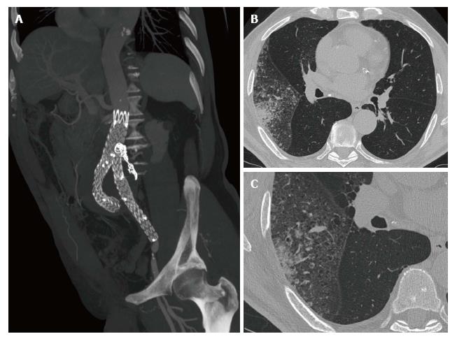
Figure 1 Lepidic predominant adenocarcinoma diagnosed during endovascular aortic aneurysm repair follow-up.
A-C: A 80-year-old male with a LPA of the right upper lobe diagnosed during endovascular aortic aneurysm repair follow-up for a type II endoleak treated with glue and coils (A). HRCT images (B and C) demonstrate the lepidic growth of the tumor and aerogenous metastases in the same lobe. LPA: Lepidic predominant adenocarcinoma; HRCT: High resolution computed tomography.
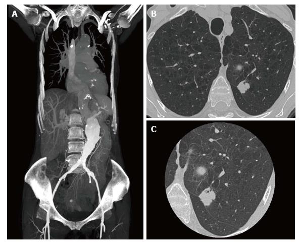
Figure 2 Breast cancer lung metastasis diagnosed during endovascular aortic aneurysm repair planning.
A-C: A-63-year-old woman, with a history of breast cancer (10 year before, pT1cN0M0), addressed to our institution for vascular planning due to an abdominal aortic aneurism (A). HRCT image (B) performed before the contrast media administration showed diffuse enphysema in upper lobes and the presence in the left upper lobe of a solid nodule (18 mm) with spiculated margins and bronchus sign, confirmed at small FOV reconstruction (C). Histological evaluation, after surgical intervention, demonstrated a breast cancer lung metastasis. HRCT: High resolution computed tomography.
- Citation: Mazzei MA, Guerrini S, Gentili F, Galzerano G, Setacci F, Benevento D, Mazzei FG, Volterrani L, Setacci C. Incidental extravascular findings in computed tomographic angiography for planning or monitoring endovascular aortic aneurysm repair: Smoker patients, increased lung cancer prevalence? World J Radiol 2017; 9(7): 304-311
- URL: https://www.wjgnet.com/1949-8470/full/v9/i7/304.htm
- DOI: https://dx.doi.org/10.4329/wjr.v9.i7.304


