Copyright
©The Author(s) 2017.
World J Radiol. Jun 28, 2017; 9(6): 269-279
Published online Jun 28, 2017. doi: 10.4329/wjr.v9.i6.269
Published online Jun 28, 2017. doi: 10.4329/wjr.v9.i6.269
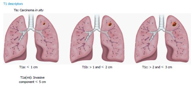
Figure 1 Illustrations demonstrating T1 descriptors.
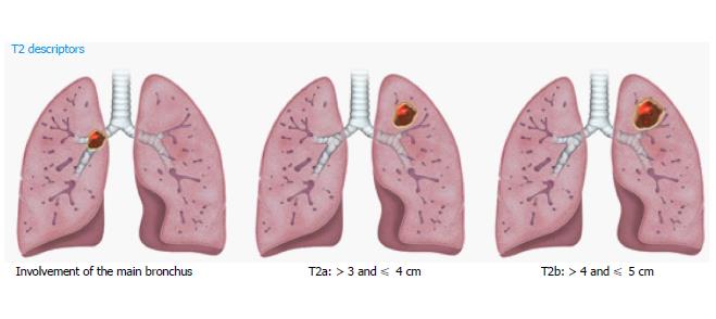
Figure 2 Illustrations demonstrating T2 descriptors.
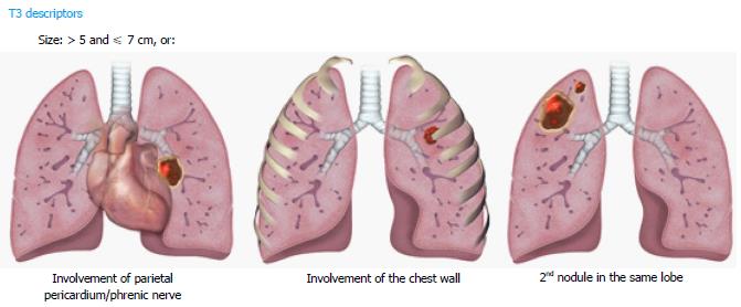
Figure 3 Illustrations demonstrating T3 descriptors.
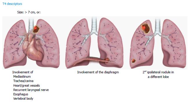
Figure 4 Illustrations demonstrating T4 descriptors.
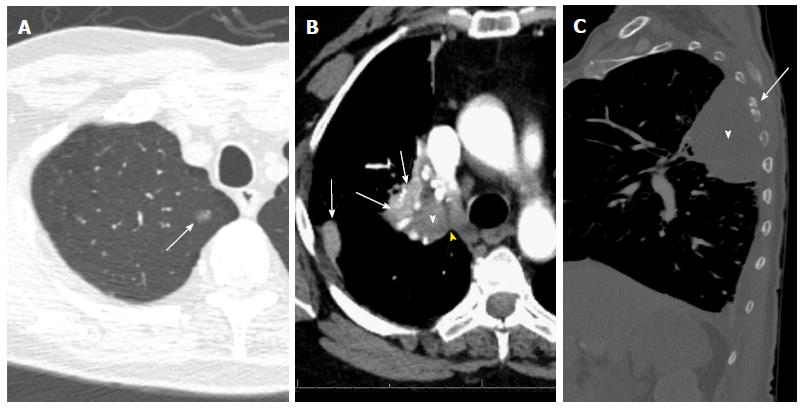
Figure 5 T descriptors.
A: T1a (mi): Groundglass opacity in the apical segment of the right upper lobe (arrow) on CT lung window image; resection revealed predominantly lepidic adenocarcinoma with 3-mm invasive component and clear margins; B: T2a: 4-cm invasive squamous cell bronchogenic carcinoma of the right upper lobar bronchus (white arrowhead) with adjacent areas of atelectasis (arrows) and clear cleavage plane with the mediastinum (yellow arrowhead) on contrast-enhanced CT image; C: T4 vs T3: 8.3cm left upper lobe mass (white arrowhead), with chest wall invasion causing destruction of the adjacent left 5th rib (arrow) and small left pleural effusion. Note that the highest descriptor should be used for T staging.
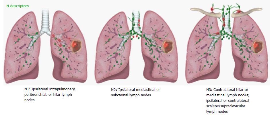
Figure 6 Illustrations demonstrating N descriptors.
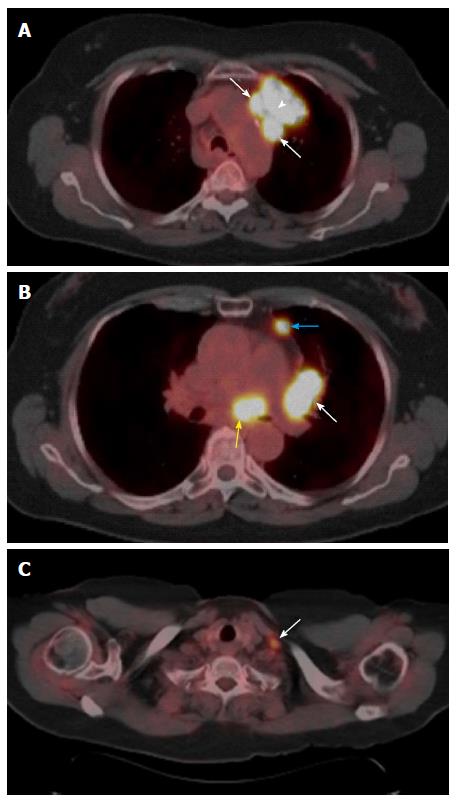
Figure 7 N descriptors.
A: Primary tumor and N2: FDG-avid left upper lobe lung mass (arrowhead) invading the prevascular region of the mediastinum with adjacent FDG-avid level 6 lymph nodes suggesting nodal metastases (arrows); B: N1 and N2: Level 10L (white arrow), and 3A (blue arrow) enlarged lymph nodes with increased FDG avidity (N1 nodal metastases) and similar level 7 (yellow arrow) lymph nodes suggesting N2 nodal metastases; C: N3: FDG-avid left supraclavicular lymph node (arrow) suggesting N3 nodal metastases.
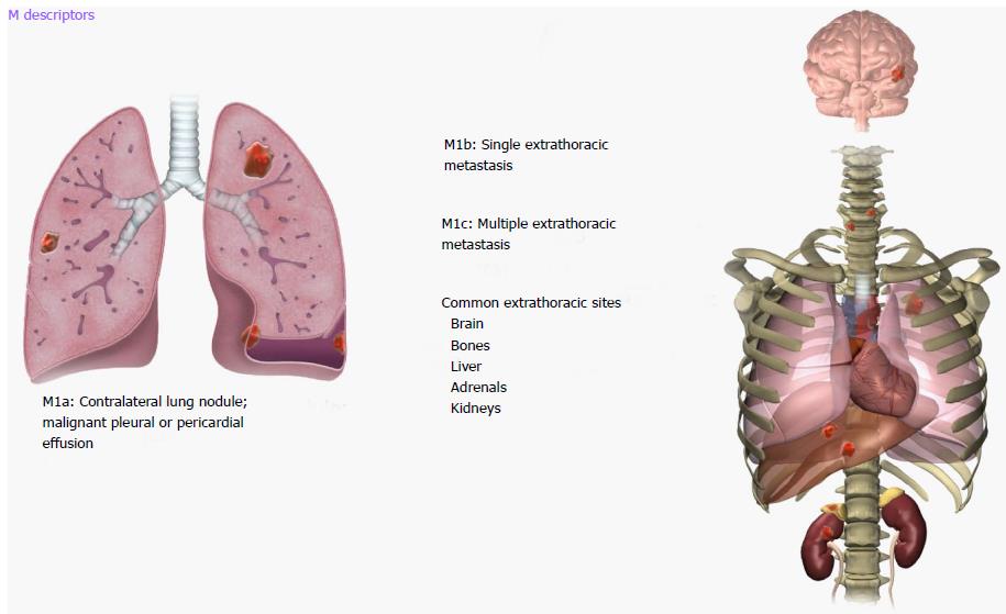
Figure 8 Illustrations demonstrating M descriptors.
Figure 9 M descriptors.
A: M1a: Nodular large heterogeneously enhancing right pleural mass like thickening (arrows) with mediastinal invasion (black arrowheads) and small right pleural effusion (white arrowhead) due to metastatic NSCLC; B: M1c: Two sclerotic metastases to thoracic spine (T3 and T9, arrows) from lung cancer; C: M1c: Multiple extrathoracic lung cancer metastases to the retroperitoneum (yellow arrows), right adrenal gland (white arrow), and spleen (blue arrow). Note the presence of malignant ascites (yellow arrowhead).
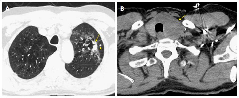
Figure 10 Challenging cases.
A: A 65-year-old man with a non-calcified spiculated lesion in the left upper lobe in the region of confluent pulmonary emphysema. The lesion reveals markedly irregular shape, with the solid component measuring 2.5 cm on computed tomography (CT) (arrow), which would put the lesion in the cT1c category. A separate solid nodule in the same lobe (arrowhead) would upstage the tumor to cT3; B: CT images at the cervicothoracic junction revealed large left thyroid lobe mass (arrow). The analysis of the left lung upper lobectomy specimen revealed a 3.2 cm primary lung adenocarcinoma, with the second nodule in the thyroid representing a metastatic thyroid cancer (pT2a lung cancer).
- Citation: Kay FU, Kandathil A, Batra K, Saboo SS, Abbara S, Rajiah P. Revisions to the Tumor, Node, Metastasis staging of lung cancer (8th edition): Rationale, radiologic findings and clinical implications. World J Radiol 2017; 9(6): 269-279
- URL: https://www.wjgnet.com/1949-8470/full/v9/i6/269.htm
- DOI: https://dx.doi.org/10.4329/wjr.v9.i6.269


