
















©The Author(s) 2017.
World J Radiol. Jun 28, 2017; 9(6): 269-279
Published online Jun 28, 2017. doi: 10.4329/wjr.v9.i6.269
Published online Jun 28, 2017. doi: 10.4329/wjr.v9.i6.269
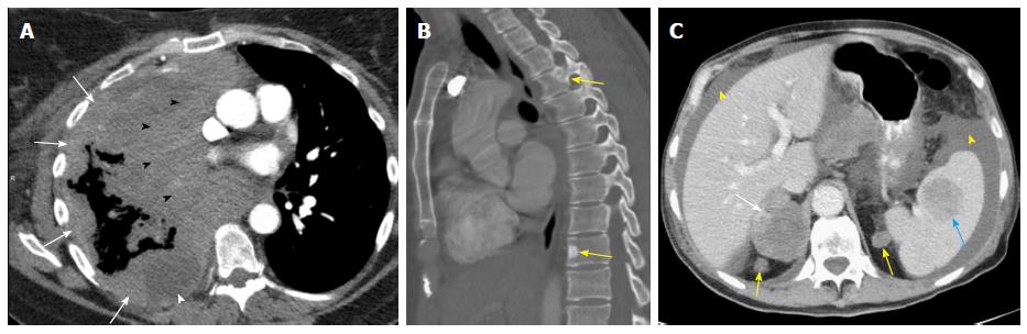
Figure 9 M descriptors.
A: M1a: Nodular large heterogeneously enhancing right pleural mass like thickening (arrows) with mediastinal invasion (black arrowheads) and small right pleural effusion (white arrowhead) due to metastatic NSCLC; B: M1c: Two sclerotic metastases to thoracic spine (T3 and T9, arrows) from lung cancer; C: M1c: Multiple extrathoracic lung cancer metastases to the retroperitoneum (yellow arrows), right adrenal gland (white arrow), and spleen (blue arrow). Note the presence of malignant ascites (yellow arrowhead).
- Citation: Kay FU, Kandathil A, Batra K, Saboo SS, Abbara S, Rajiah P. Revisions to the Tumor, Node, Metastasis staging of lung cancer (8th edition): Rationale, radiologic findings and clinical implications. World J Radiol 2017; 9(6): 269-279
- URL: https://www.wjgnet.com/1949-8470/full/v9/i6/269.htm
- DOI: https://dx.doi.org/10.4329/wjr.v9.i6.269