Copyright
©The Author(s) 2017.
World J Radiol. Apr 28, 2017; 9(4): 148-154
Published online Apr 28, 2017. doi: 10.4329/wjr.v9.i4.148
Published online Apr 28, 2017. doi: 10.4329/wjr.v9.i4.148
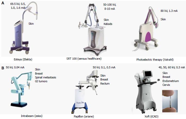
Figure 1 Electronic brachytherapy systems operating.
A: The range between 50 and 100 kVp; B: The range between 40 and 60 kVp.
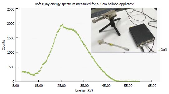
Figure 2 Xoft X-ray energy spectrum measured using an Amptek spectrometer.
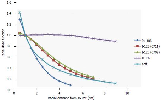
Figure 3 Radial dose function [Pd-103, I-125 (6711), I-125 (6702), Ir-192 and Xoft].
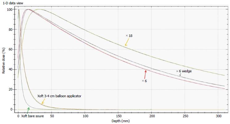
Figure 4 Comparison of 6MV, 6MV wedge, 18 MV, Xoft bare source and Xoft 3-4 cm balloon applicator profiles.
Figure 5 Simulation of a Tandem and Ovoid applicators planned in a Brachyvision software for a predefined Xoft X-ray source dwell positions.
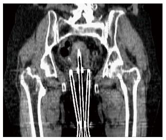
Figure 6 Coronal view of the Xoft Henschke applicator.
- Citation: Ramachandran P. New era of electronic brachytherapy. World J Radiol 2017; 9(4): 148-154
- URL: https://www.wjgnet.com/1949-8470/full/v9/i4/148.htm
- DOI: https://dx.doi.org/10.4329/wjr.v9.i4.148


