Copyright
©The Author(s) 2017.
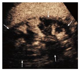
Figure 1 Patient with solitary kidney developing acute renal failure.
Contrast enhanced ultrasound showed multiple renal infarctions (arrows) involving a large portion of the parenchyma.
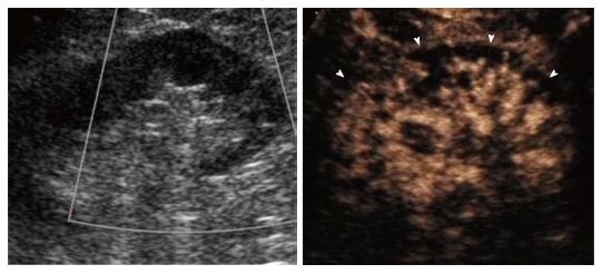
Figure 2 Patient with grade III chronic renal failure developing acute renal failure after Endovascular aortic repair.
A: Color Doppler ultrasound showed avascular kidney; B: Contrast enhanced ultrasound showed enhancing hilar vessels and lack of enhancement of large portions of the cortex (arrowheads) consistent with acute cortical necrosis. The contralateral kidney was normal (not shown).
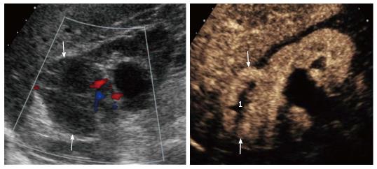
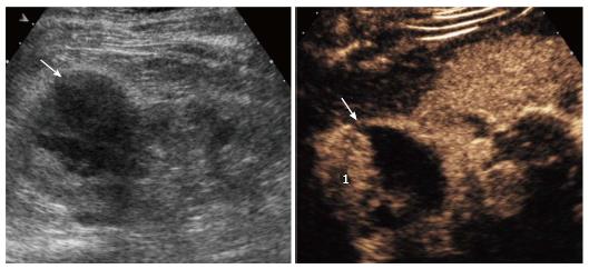
Figure 3 Patient with grade III chronic renal failure.
A: Color Doppler ultrasound showed markedly reduced renal parenchyma perfusion and a hypoechoic lesion without obvious vascularity (arrows); B: Contrast enhanced ultrasound showed a solid enhancing mass (arrows) with avascular central portion (1). A clear cell RCC with necrotic central areas was found at surgery. RCC: Renal cell carcinoma.
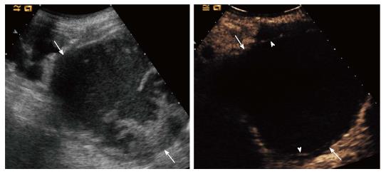
Figure 4 Patient with grade IV chronic renal failure.
A: Grey-scale ultrasound showed a complex renal lesion (arrows). Contrast enhanced ultrasound showed no intralesional enhancement, nor vegetations; B: Two thin septa were visible (arrowheads) with minimum enhancement (benign minimally complicated cysts, Bosniak category II).
Figure 5 Patient with grade III chronic renal failure that one category IV lesion was a clear cell renal cell carcinoma at nephrectomy.
A: Grey-scale ultrasound showed a complex renal lesion (arrow); B: Contrast enhanced ultrasound showed thickened wall with a vegetation (1) consistent for a presumably malignant, Bosniak category IV lesion. A clear cell renal cell carcinoma was found at surgery.
- Citation: Girometti R, Stocca T, Serena E, Granata A, Bertolotto M. Impact of contrast-enhanced ultrasound in patients with renal function impairment. World J Radiol 2017; 9(1): 10-16
- URL: https://www.wjgnet.com/1949-8470/full/v9/i1/10.htm
- DOI: https://dx.doi.org/10.4329/wjr.v9.i1.10


