Copyright
©The Author(s) 2017.
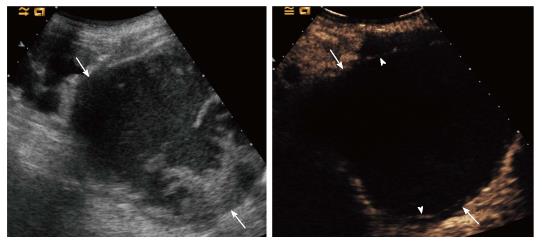
Figure 4 Patient with grade IV chronic renal failure.
A: Grey-scale ultrasound showed a complex renal lesion (arrows). Contrast enhanced ultrasound showed no intralesional enhancement, nor vegetations; B: Two thin septa were visible (arrowheads) with minimum enhancement (benign minimally complicated cysts, Bosniak category II).
- Citation: Girometti R, Stocca T, Serena E, Granata A, Bertolotto M. Impact of contrast-enhanced ultrasound in patients with renal function impairment. World J Radiol 2017; 9(1): 10-16
- URL: https://www.wjgnet.com/1949-8470/full/v9/i1/10.htm
- DOI: https://dx.doi.org/10.4329/wjr.v9.i1.10


