Copyright
©The Author(s) 2017.
Figure 1 Native T1-map, post-contrast T1-map, and extracellular volume map of the myocardium in a healthy subject.
ECV: Extracellular volume.
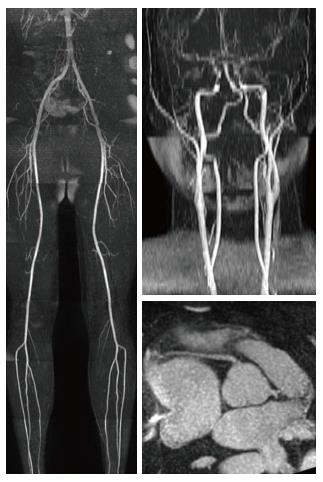
Figure 2 Visualization of the lower extremity, carotid and coronary arteries using quiescent-interval single-shot magnetic resonance angiography.
- Citation: De Cecco CN, Muscogiuri G, Varga-Szemes A, Schoepf UJ. Cutting edge clinical applications in cardiovascular magnetic resonance. World J Radiol 2017; 9(1): 1-4
- URL: https://www.wjgnet.com/1949-8470/full/v9/i1/1.htm
- DOI: https://dx.doi.org/10.4329/wjr.v9.i1.1


