Copyright
©The Author(s) 2016.
World J Radiol. May 28, 2016; 8(5): 513-517
Published online May 28, 2016. doi: 10.4329/wjr.v8.i5.513
Published online May 28, 2016. doi: 10.4329/wjr.v8.i5.513
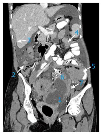
Figure 1 In a 67-year-old patient with advanced stage ovarian carcinoma, the post-contrast computed tomography coronal image depicts preferential spread of peritoneal implant involvement of rectouterine pouch (1), right paracolic gutter (2), right perihepatic space (3), inframesocolon (4), left paracolic gutter (5), mesentery (6) and mesosigmoid (7).
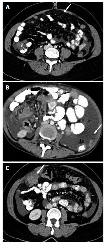
Figure 2 Computed tomography image showing peritoneal and omental deposits in ovarian carcinoma.
Subtle stranding (A), nodule (B) and mass like deposit (C) are three forms of peritoneal involvement.
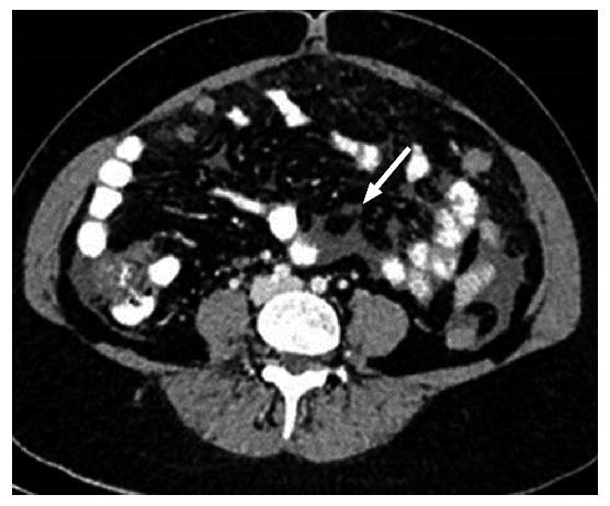
Figure 3 Axial contrast-enhanced computed tomography image shows a small nodule in the mesentery.
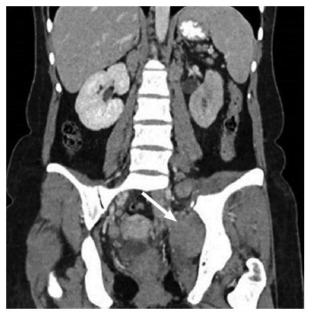
Figure 4 Coronal post-contrast computed tomography image shows pelvic side wall involvement.
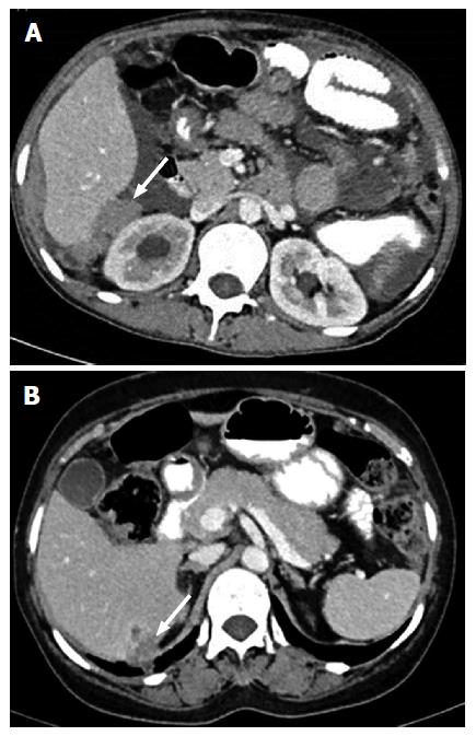
Figure 5 Contrast-enhanced computed tomography in two different patients showing deposits in Morrison’s pouch (A) and liver parenchyma invasion by surface deposit (B).
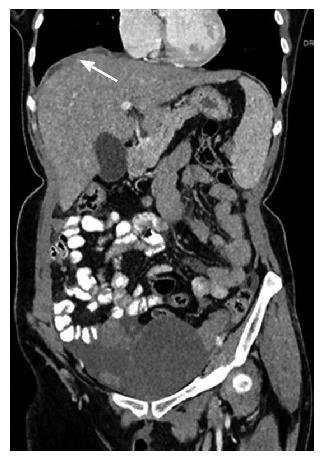
Figure 6 Coronal image showing smooth minimal peritoneal thickening in right subphrenic space.
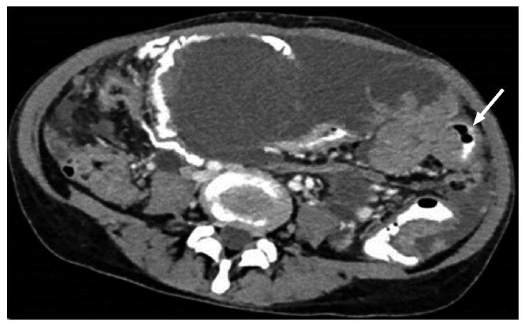
Figure 7 Axial computed tomography showing smooth, diffuse, eccentric bowel wall thickening involving one of the small bowel loops.
Mild thickening best seen in oral contrast distended bowel loop.
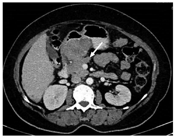
Figure 8 Solid nodular deposit in lesser omentum (gastrohepatic ligament) in a 40-year-old female patient with stage III ovarian carcinoma.
Figure 9 Lobulated mass like deposit in lesser sac in a case of advanced ovarian carcinoma.
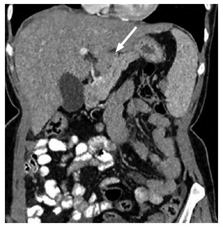
Figure 10 III-defined soft tissue along falciform ligament in a 50-year-old patient with ovarian carcinoma.
It has to be differentiated from liver parenchyma lesion.
- Citation: Chandrashekhara SH, Triveni GS, Kumar R. Imaging of peritoneal deposits in ovarian cancer: A pictorial review. World J Radiol 2016; 8(5): 513-517
- URL: https://www.wjgnet.com/1949-8470/full/v8/i5/513.htm
- DOI: https://dx.doi.org/10.4329/wjr.v8.i5.513


