
















©The Author(s) 2016.
World J Radiol. May 28, 2016; 8(5): 513-517
Published online May 28, 2016. doi: 10.4329/wjr.v8.i5.513
Published online May 28, 2016. doi: 10.4329/wjr.v8.i5.513
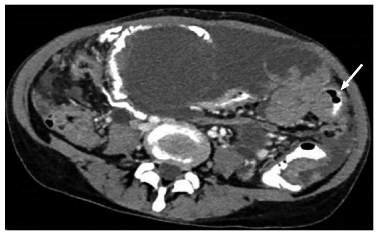
Figure 7 Axial computed tomography showing smooth, diffuse, eccentric bowel wall thickening involving one of the small bowel loops.
Mild thickening best seen in oral contrast distended bowel loop.
- Citation: Chandrashekhara SH, Triveni GS, Kumar R. Imaging of peritoneal deposits in ovarian cancer: A pictorial review. World J Radiol 2016; 8(5): 513-517
- URL: https://www.wjgnet.com/1949-8470/full/v8/i5/513.htm
- DOI: https://dx.doi.org/10.4329/wjr.v8.i5.513