Copyright
©The Author(s) 2016.
World J Radiol. Apr 28, 2016; 8(4): 390-396
Published online Apr 28, 2016. doi: 10.4329/wjr.v8.i4.390
Published online Apr 28, 2016. doi: 10.4329/wjr.v8.i4.390
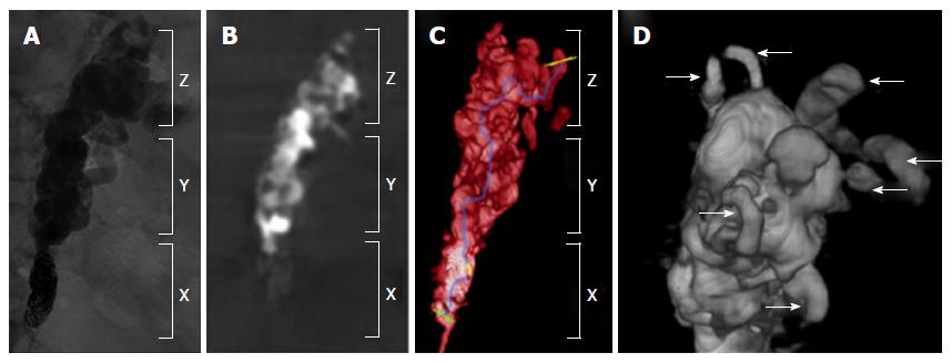
Figure 1 A radiographic illustration of cone-beam computed tomography during the modified balloon-occluded retrograde transvenous obliteration procedure.
A: Fluoroscopic image of CARTO demonstrating coil blockage (X), gastrorenal shunt (Y) and gastric varices (Z). As shown, it is difficult to assess the degree of obliteration/thrombosis of the gastric varices on 2D fluoroscopic image; B: Selective 2D reconstructed CBCT image of mBRTO corresponding to the fluoroscopic image and (C) 3D reconstructed CBCT image of mBRTO providing additional information of an amount of obliteration, a course of gastrorenal shunt and numerous collaterals. This confirmed the complete obliteration of gastric varices including collaterals; D: An enlarged view of 3D reconstructed CBCT image of gastric varices demonstrating multiple collateral afferent and efferent collaterals (white arrows). CBCT: Cone-beam computed tomography; mBRTO: Modified balloon-occluded retrograde transvenous obliteration; CARTO: Coil-assisted retrograde transvenous obliteration.
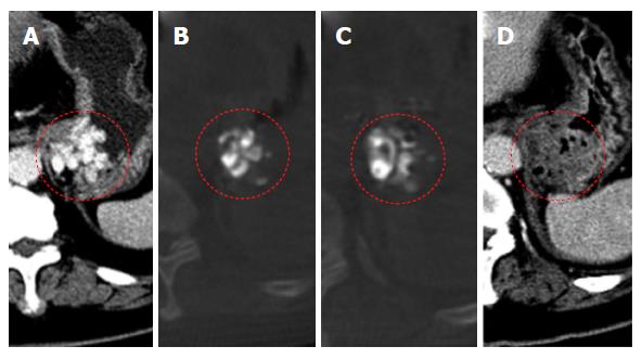
Figure 2 Selective contrast-enhanced portal-venous phase axial computed tomography image.
A: Selective contrast-enhanced portal-venous phase axial CT image of gastric varices in (A) pre-mBRTO with multiple transmural gastric varices in the gastric fundus, (B and C) selective 2D reconstructed CBCT images demonstrating multiple gelfoam/contrast filled gastric varices at a different slice confirming complete obliteration of corresponding gastric varices shown in (A). D: Selective contrast enhanced portal venous phase axial CT image 2-d post-mBRTO demonstrating non-enhancing/hypoenhancing gastric fundal wall consisted with completely thrombosed gastric varices consistent with findings of CBCT images. CT: Computed tomography; CBCT: Cone-beam CT; mBRTO: Modified balloon-occluded retrograde transvenous obliteration.
- Citation: Lee EW, So N, Chapman R, McWilliams JP, Loh CT, Busuttil RW, Kee ST. Usefulness of intra-procedural cone-beam computed tomography in modified balloon-occluded retrograde transvenous obliteration of gastric varices. World J Radiol 2016; 8(4): 390-396
- URL: https://www.wjgnet.com/1949-8470/full/v8/i4/390.htm
- DOI: https://dx.doi.org/10.4329/wjr.v8.i4.390


