Copyright
©The Author(s) 2016.
World J Radiol. Apr 28, 2016; 8(4): 378-389
Published online Apr 28, 2016. doi: 10.4329/wjr.v8.i4.378
Published online Apr 28, 2016. doi: 10.4329/wjr.v8.i4.378
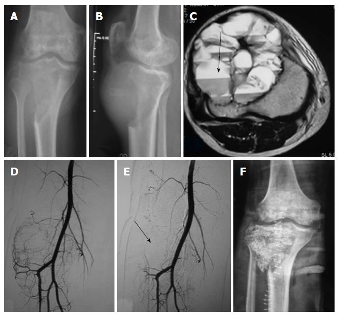
Figure 1 Preoperative embolisation of giant cell tumor of upper end of tibia.
A-F: Antero-posterior (A) and lateral (B) plain radiographs of right knee show expansile lytic destructive lesion involving proximal end of right tibia reaching up to subarticular location suggestive of giant cell tumor; Axial (C) T2-weighted fat saturated magnetic resonance image show heterogeneously hyperintense lesion with blood-fluid levels within it suggestive of secondary aneurysmal bone cysts formation (arrow); Pre embolisation non-selective DSA (D) image of left SFA show multiple feeders arising from distal SFA and proximal anterior tibial artery supplying the tumor with marked tumor blush; Post embolisation non-selective DSA (E) image show > 90% reduction in tumor blush (arrow); Postoperative AP plain radiograph of right knee (F) show replacement of bony defect following tumor curettage with combined autograft (iliac crest) and allograft. DSA: Digital substraction angiography; SFA: Superficial femoral artery.
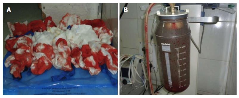
Figure 2 Photograph of gauze pieces and suction bottle used to measure blood loss during the study.
Operative photograph (A) show blood soaked gauze pieces that were weighed to calculate blood loss and photograph of suction bottle (B) show collected blood during surgery.
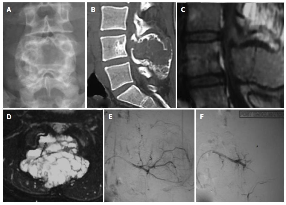
Figure 3 Preopertaive embolisation of spinal aneurysmal bone cyst.
Antero-posterior (A) plain radiograph of lumbo-sacral spine show expansile lytic lesion involving the posterior elements of L4 vertebra suggestive of aneurysmal bone cyst; Sagittal (B) 3D-reformatted non-contrast computed tomography of lumbo-sacral spine show lytic expansile destructive lesion involving posterior elements of L4 vertebra; Sagittal (C) and axial (D) T2-weighted fat saturated magnetic resonance images show T2 hyperintense lesion involving posterior element of L4 vertebra with blood-fluid levels (arrow); Pre embolisation non-selective digital substraction angiography (DSA) (E) image of L3 lumbar artery showmultiple feeders supplying the tumor with tumor blush; Post embolisation DSA (F) image show more than 90% reduction in tumor blush (asterix).
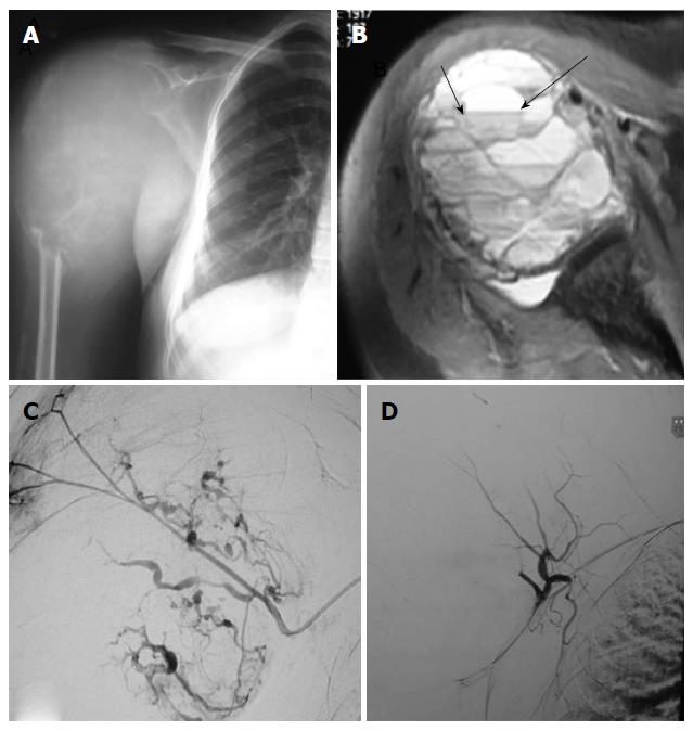
Figure 4 Preoperative embolisation of aneurysmal bone cyst of humerus.
Antero-posterior (A) plain radiograph of right shoulder show expansile lytic destructive lesion involving proximal end ofright humerus, suggestive of aneurysmal bone cyst; Axial (B) T2-weighted fat suppressed magnetic resonance image showlesion is hyperintense with fluid-fluid levels within it (arrow); Digital substraction angiography (C) image of right circumflex humeral artery show multiple feeders supplying the tumor with marked tumor blush; Post embolisation angiogram (D) of circumflex humeral artery show near complete reduction of tumor blush.
- Citation: Jha R, Sharma R, Rastogi S, Khan SA, Jayaswal A, Gamanagatti S. Preoperative embolization of primary bone tumors: A case control study. World J Radiol 2016; 8(4): 378-389
- URL: https://www.wjgnet.com/1949-8470/full/v8/i4/378.htm
- DOI: https://dx.doi.org/10.4329/wjr.v8.i4.378


