Copyright
©The Author(s) 2016.
World J Radiol. Mar 28, 2016; 8(3): 268-274
Published online Mar 28, 2016. doi: 10.4329/wjr.v8.i3.268
Published online Mar 28, 2016. doi: 10.4329/wjr.v8.i3.268
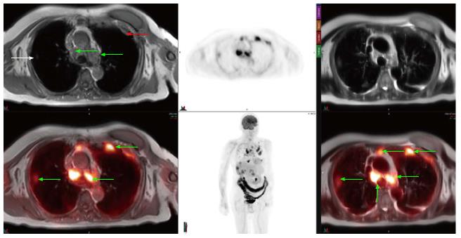
Figure 1 A 65-year-old patient with osteosarcoma underwent positron emission tomography-magnetic resonance imaging for annual follow-up.
There were multiple nodular T1 isointense and T2 hyperintense foci (relative to skeletal muscle) noted which demonstrated FDG uptake, compatible with neoplastic activity. Biopsy were positive for metastatic osteosarcoma. FDG: 18F-fluorodeoxyglucose.
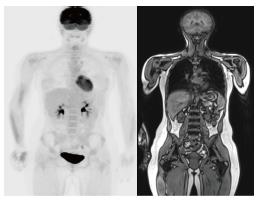
Figure 2 A 17-year-old male with history of Ewing sarcoma underwent follow up whole body positron emission tomography-magnetic resonance imaging which did not reveal any focal abnormal 18F-fluorodeoxyglucose uptake nor was there any focal abnormal marrow signal within the imaged musculoskeletal system to suggest recurrence.
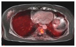
Figure 3 A 63-year-old female with renal failure and abnormal serum protein electrophoresis.
Positron emission tomography-magnetic resonance imaging revealed focal abnormal heterogeneously T1 hypointense and T2 hyperintense lesion with corresponding increased FDG uptake. Lesion biopsy was positive for multiple myeloma. FDG: 18F-fluorodeoxyglucose.
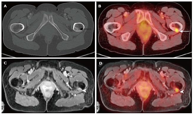
Figure 4 A 59-year-old female with history of breast cancer status post resection and chemoradiation presents with left hip pain.
Noncontrast CT of the pelvis (A) was unremarkable. PET/CT revealed increased focus of FDG uptake within the proximal left femur. Contrast-enhanced pelvic MRI (C) and PET-MRI (D) acquired simultaneously demonstrates abnormal nodular soft tissue mass within the proximal left femoral cortex with increased radiotracer uptake compatible with metastasis in this patient with breast carcinoma. PET: Positron emission tomography; MRI: Magnetic resonance imaging; CT: Computed tomography; FDG: 18F-fluorodeoxyglucose.
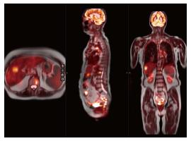
Figure 5 A 66-year-old female with history of breast cancer status post resection and chemoradiation presents with chest pain.
Whole-body PET-MRI reveal multiple abnormal nodular soft tissue masses within the lungs, mediastinum, ribs, liver and lumbar spine with increased FDG uptake consistent with metastasis in this patient with breast carcinoma. PET: Positron emission tomography; MRI: Magnetic resonance imaging; FDG: 18F-fluorodeoxyglucose.
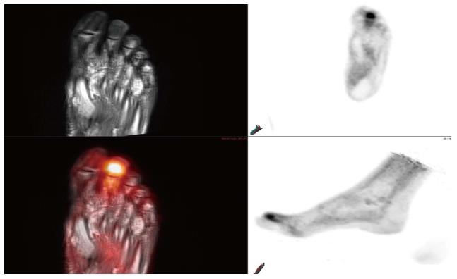
Figure 6 A 71-year-old male with poorly controlled diabetes and end-stage renal disease presents with worsening right foot pain.
Bone scan revealed diffuse radiotracer uptake within the right foot without focal abnormality. MRI of the right foot demonstrate diffuse abnormal T1 and T2 signal within the right foot digits. Multiplanar PET-MRI images reveal focal FDG uptake within the distal right second digit with corresponding heterogeneous T1 hypointense, T2 hyperintense signal compatible with osteomyelitis. PET: Positron emission tomography; MRI: Magnetic resonance imaging; FDG: 18F-fluorodeoxyglucose.
- Citation: Chaudhry AA, Gul M, Gould E, Teng M, Baker K, Matthews R. Utility of positron emission tomography-magnetic resonance imaging in musculoskeletal imaging. World J Radiol 2016; 8(3): 268-274
- URL: https://www.wjgnet.com/1949-8470/full/v8/i3/268.htm
- DOI: https://dx.doi.org/10.4329/wjr.v8.i3.268


