Copyright
©The Author(s) 2015.
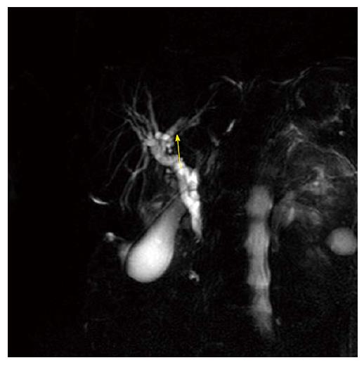
Figure 1 Little stones in left branch of biliary tree (arrow).
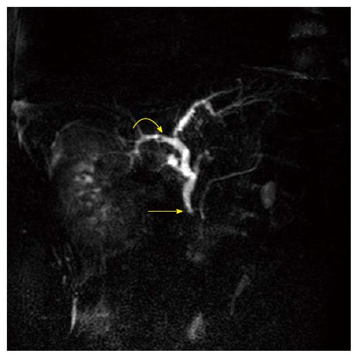
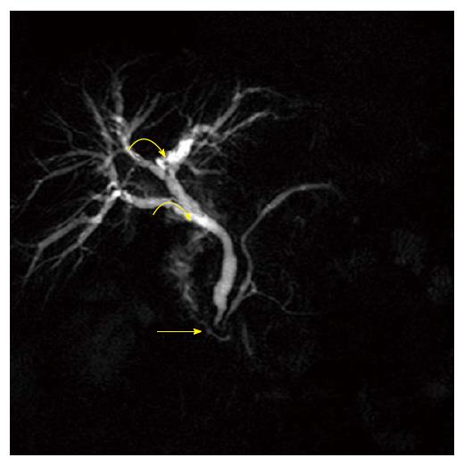
Figure 2 Pre-papillary micro stones (arrow) fake stone in right branch of biliary tree (curved arrow).
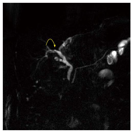
Figure 3 (Same patient of Figure 2) next image shows the fake stone as biliary branches crossing (curved arrow).
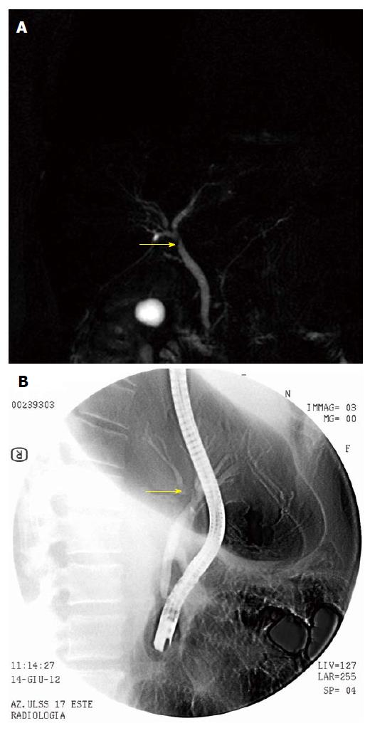
Figure 4 Stricture or stones (arrow) at origin of common bile duct (A) and stricture (arrow) confirmed at endoscopic retrograde cholangiopancreatography but the balloon-catheter pulled out micro stones and no stricture was confirmed at control (B).
Figure 5 Pre-papillary stricture (arrow) microstones in right and left branch (curved arrows, confirmed at endoscopic retrograde cholangiopancreatography).
Figure 6 Micro stones in gallbladder (curved arrow), pre-papillary stenosis (arrow) (A) and endoscopic retrograde cholangiopancreatography shows pre-papillary stone (arrow), not the micro stones that were pulled out by balloon-catheter (B).
- Citation: Polistina FA, Frego M, Bisello M, Manzi E, Vardanega A, Perin B. Accuracy of magnetic resonance cholangiography compared to operative endoscopy in detecting biliary stones, a single center experience and review of literature. World J Radiol 2015; 7(4): 70-78
- URL: https://www.wjgnet.com/1949-8470/full/v7/i4/70.htm
- DOI: https://dx.doi.org/10.4329/wjr.v7.i4.70


