Copyright
©2014 Baishideng Publishing Group Inc.
World J Radiol. Aug 28, 2014; 6(8): 567-582
Published online Aug 28, 2014. doi: 10.4329/wjr.v6.i8.567
Published online Aug 28, 2014. doi: 10.4329/wjr.v6.i8.567
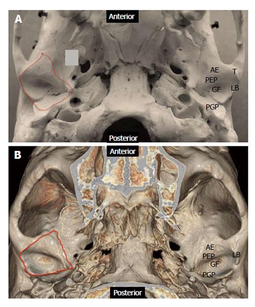
Figure 1 Anatomy of the cranial component of temporomandibular joint.
A: Photograph of skull specimen; B: 3-D volume rendered image obtained from a temporal bone Redline demonstrates the capsular attachment. AE: Articular eminence; GF: Glenoid fossa; LB: Lateral border; PEP: Preglenoid plane; PGP: Postglenoid plane; T: Tubercle.
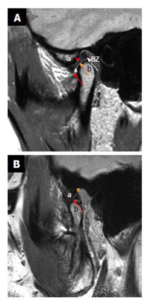
Figure 2 Normal anatomy.
Sagittal proton density weighted closed mouth and open mouth view of magnetic resonance imaging. A: On the closed mouth view, the disk is located posterior to the articular eminence (the letter, a). It can be noted that the “bow-tie” shape of the disk: Thicker anterior band (red arrow) and posterior band (white arrow) with a thinner central zone (orange arrow). Bilaminar zone (BZ) is located posterior to the posterior band. It can also be noted that the inferior joint compartment (white arrowhead) between the disk and the mandibular condyle (the letter, b) and superior joint compartment (red arrowhead) between the articular eminence and the disk; B: On the open mouth view (in a different patient), the thinner intermediate zone (red arrow) of the disk is interposed between the articular eminence (the letter, a) and the condylar head (the letter, b) in a “bow-tie” fashion. Orange arrowhead demonstrates temporal lamina and black arrowhead indicate inferior lamina.
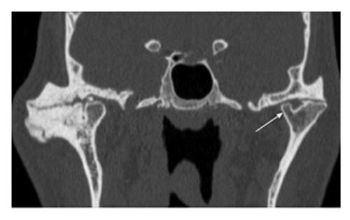
Figure 3 Bifid condyle.
Coronal reformatted computed tomography image through the temporomandibular joint (TMJ) demonstrates bifid left mandibular condyle. It can be noted that one of the condyles (arrow) is smaller than the other. Advanced degenerative changes are noted in bilateral TMJ.
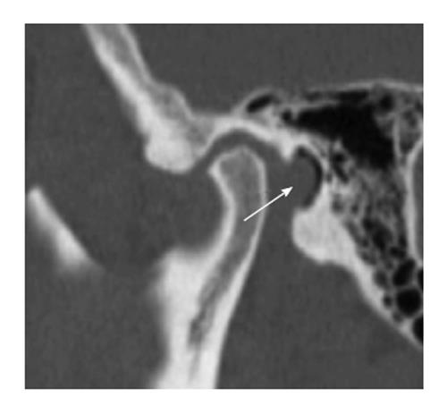
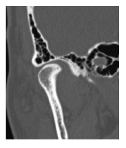
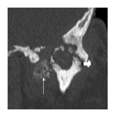
Figure 4 Foramen of Huschke.
Sagittal reformatted computed tomography image through the temporomandibular joint demonstrates a focal defect (arrow) in the tympanic plate.
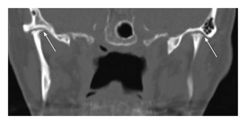
Figure 5 Idiopathic condylar resorption.
Coronal reformatted computed tomography image through the temporomandibular joint of a young patient demonstrates bilateral severe condylar resorption (arrows) without any evidence of degenerative changes within the joint.
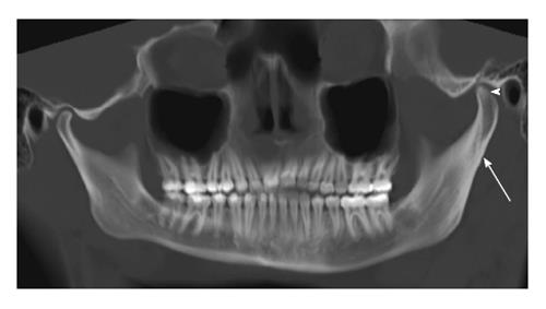
Figure 6 Condylar hyperplasia.
Panoramic reformation of the source computed tomography data including both the temporomandibular joints of a young patient demonstrates hyperplasia of the left condyle (arrowhead) in comparison to the right side. Associated hypertrophy of the ramus and the neck (arrow) of the left hemi-mandible is also noted.
Figure 7 Extensive pneumatization.
Coronal reformatted computed tomography image through the right temporomandibular joint demonstrates almost complete pneumatization of the glenoid fossa except the central part.
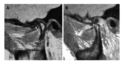
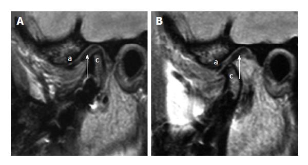
Figure 8 Anterior displacement with reduction.
A: Sagittal proton density weighted magnetic resonance imaging (MRI) in the closed mouth position demonstrates anterior displacement of the disk (arrow) in front of the mandibular condyle (the letter, c); B: Sagittal proton density weighted MRI in the open mouth position demonstrates reduction of the disk (arrow) between the articular eminence (the letter, a) and the mandibular condyle (the letter, c).
Figure 9 Anterior displacement with no reduction.
A: Sagittal proton density weighted magnetic resonance imaging (MRI) in the closed mouth position demonstrates anterior displacement of the disk (arrow) related to the articular eminence (the letter, a) and anterior to the mandibular condyle (the letter c); B: Sagittal proton density weighted MRI in the open mouth position demonstrates no reduction of the disk (arrow) between the articular eminence (the letter, a) and the mandibular condyle (the letter, c).
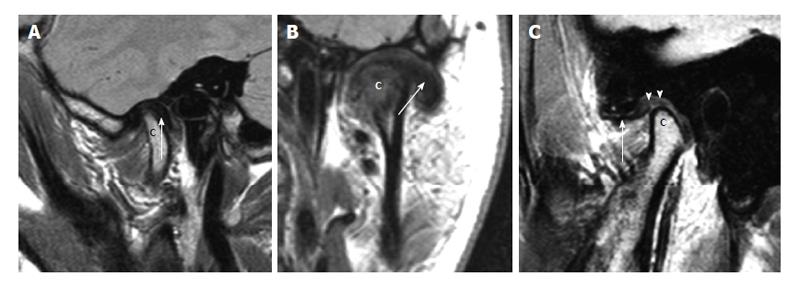
Figure 10 Other types of disk displacement.
A: Posterior disk displacement. Sagittal proton density weighted magnetic resonance imaging (MRI) in the closed mouth position demonstrates posterior displacement of the disk (arrow) in relation to the mandibular condyle (the letter, c); B: Lateral disk displacement. Coronal proton density weighted demonstrates lateral displacement of the disk (arrow) in relation to the mandibular condyle (the letter, c); C: Pseudodisk. Sagittal proton density weighted MRI in the closed mouth position demonstrates anterior displacement of the disk (arrow) in front of the mandibular condyle (the letter, c). The thickening of the posterior attachments (arrowheads) superior to the mandibular condyle is seen as “pseudodisk”.
Figure 11 Stuck disk.
A: Sagittal proton density weighted magnetic resonance imaging (MRI) in the closed mouth position demonstrates apparently normal position of the disk (arrow) in relation to the mandibular condyle (the letter, c). The letter “a” demonstrates the articular eminence; B: Sagittal proton density weighted MRI in the open mouth position demonstrates no anterior movement of the disk (arrow) with the mandibular condyle (the letter, c), i.e., “stuck” to the glenoid fosssa. The articular eminence is denoted with letter “a”.
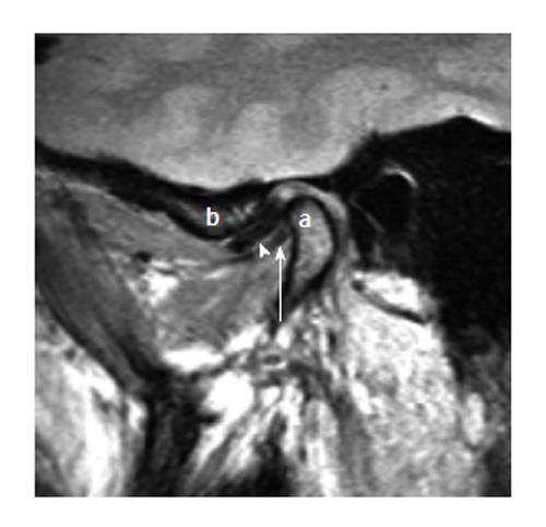
Figure 12 Double disk sign (thickening of the lateral pterygoid muscle).
Sagittal closed mouth proton density image demonstrates anterior displacement of the disk (arrow head). The thickened lateral pterygoid muscle near the mandibular condylar (the letter, a) attachment appear as linear hypointense structure (white arrow) inferior to the disk in the same orientation giving the appearance of “double disk”. The articular eminence is denoted with letter “b”.
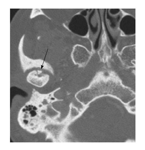
Figure 13 Osteochondritis dessicans.
Axial computed tomography scan through the level of the temporomandibular joint demonstrates a tiny bone fragment (arrow) at the anterior aspect of the disk. It can be noted that there are linear lucency surrounding the bone fragment.
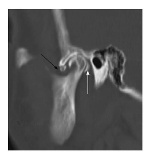
Figure 14 Loose bodies.
Sagittal reformation of the axial dataset demonstrates multiple “loose bodies” in the joint cavities, anteroinferior to the articular eminence (black arrow) and immediately posterior to the mandibular condyle (white arrow).
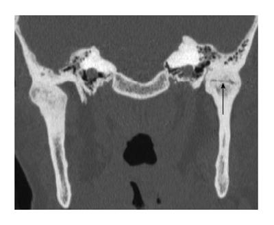
Figure 15 Ankylosis.
Coronal reformation of the axial dataset demonstrates complete ankylosis of the right temporomandibular joint (TMJ) and near complete ankylosis of the left TMJ with subtle residual joint space at the center (black arrow).
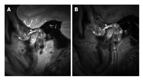
Figure 16 Juvenile idiopathic arthritis.
A: Sagittal proton density weighted magnetic resonance imaging (MRI) in the closed mouth position demonstrates increased signal at the mandibular condyle (the letter, c), extensive thickening of the synovium (the letter, s) in the retrodiscal regions. It can be noted that the thickening and increased signal of the synovium at other places (arrowheads); B: Sagittal fat suppressed post contrast T1 weighted MRI in the closed mouth position demonstrates enhancement of signal at the mandibular condyle (the letter, c), enhancement and extensive thickening of the synovium (the letter, s) in the retrodiscal regions. There is thickening and enhancement of the synovium at other places (arrowheads).
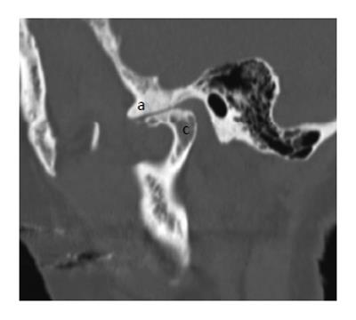
Figure 17 Degenerative changes.
Sagittal reformation of the axial dataset demonstrates deformity of the mandibular condyle (the letter, c), extensive sclerosis of the articular eminence (the letter, a) and severe loss of joint space.
Figure 18 Calcium pyrophosphate dehydrate deposition disease.
Coronal reformation of the axial dataset demonstrates destruction of the left temporomandibular joint with erosion and deformity of both the mandibular condyle and the glenoid fossa. There is extensive extensive calcium pyrophosphate dehydrate deposition disease medial to the joint space (arrow).
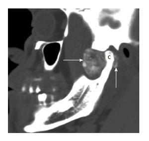
Figure 19 Synovial chondromatosis.
Sagittal reformation of the axial dataset demonstrates extensive cloud-like calcification (arrows) filling and expanding the joint space anterior to the mandibular condyle (the letter, c). Calcification is also present posterior to the mandibular condyle.
- Citation: Bag AK, Gaddikeri S, Singhal A, Hardin S, Tran BD, Medina JA, Curé JK. Imaging of the temporomandibular joint: An update. World J Radiol 2014; 6(8): 567-582
- URL: https://www.wjgnet.com/1949-8470/full/v6/i8/567.htm
- DOI: https://dx.doi.org/10.4329/wjr.v6.i8.567


