Copyright
©2014 Baishideng Publishing Group Inc.
World J Radiol. Aug 28, 2014; 6(8): 567-582
Published online Aug 28, 2014. doi: 10.4329/wjr.v6.i8.567
Published online Aug 28, 2014. doi: 10.4329/wjr.v6.i8.567
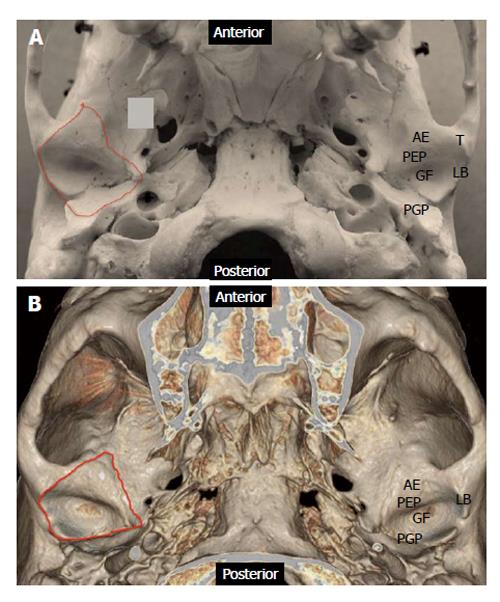
Figure 1 Anatomy of the cranial component of temporomandibular joint.
A: Photograph of skull specimen; B: 3-D volume rendered image obtained from a temporal bone Redline demonstrates the capsular attachment. AE: Articular eminence; GF: Glenoid fossa; LB: Lateral border; PEP: Preglenoid plane; PGP: Postglenoid plane; T: Tubercle.
- Citation: Bag AK, Gaddikeri S, Singhal A, Hardin S, Tran BD, Medina JA, Curé JK. Imaging of the temporomandibular joint: An update. World J Radiol 2014; 6(8): 567-582
- URL: https://www.wjgnet.com/1949-8470/full/v6/i8/567.htm
- DOI: https://dx.doi.org/10.4329/wjr.v6.i8.567


