Copyright
©2014 Baishideng Publishing Group Inc.
World J Radiol. Aug 28, 2014; 6(8): 538-543
Published online Aug 28, 2014. doi: 10.4329/wjr.v6.i8.538
Published online Aug 28, 2014. doi: 10.4329/wjr.v6.i8.538
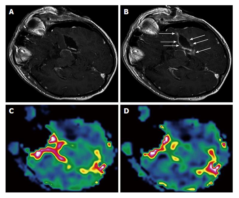
Figure 1 Surgically induced intraoperative contrast leakage.
Reprinted from NeuroImage with permission[9]. A: T1-weighted magnetic resonance (MR) image of the initial resection control. Residual tumor was depicted (not shown), neuronavigation was updated and the residual tumor was removed; B: T1-weighted MR image in identical orientation as in (A) of the second intraoperative resection control. At the border of the resection cavity there is contrast enhancement of previously non-enhancing tissue (arrows), which is caused by the neurosurgical resection leading to a leakage phenomenon. Perfusion maps of rCBV (C) and rCBF (D) at the second resection control demonstrate no elevated values in areas of contrast enhancement but complete resection of the tumor. rCBV: Regional cerebral blood volume; rCBF: Regional cerebral blood flow.
- Citation: Ulmer S. Intraoperative perfusion magnetic resonance imaging: Cutting-edge improvement in neurosurgical procedures. World J Radiol 2014; 6(8): 538-543
- URL: https://www.wjgnet.com/1949-8470/full/v6/i8/538.htm
- DOI: https://dx.doi.org/10.4329/wjr.v6.i8.538


