Copyright
©2014 Baishideng Publishing Group Inc.
World J Radiol. Jun 28, 2014; 6(6): 284-300
Published online Jun 28, 2014. doi: 10.4329/wjr.v6.i6.284
Published online Jun 28, 2014. doi: 10.4329/wjr.v6.i6.284
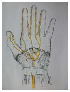
Figure 1 Sketch of the palm, showing specific details of the inner structures of the carpal tunnel (inside the wrist).
The median nerve and its branches after the wrist are marked in yellow.
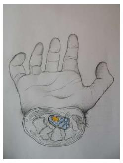
Figure 2 Sketch of the cross-section of the carpal tunnel on a hand.
Median nerve is shown in yellow and the nine flexor tendons are marked in blue.
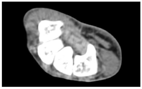
Figure 3 Axial computed tomography scan shows bony part of carpal tunnel at the level of outlet.
Bony structures from left to right are HAMATE, CAPITATE, TRAPEZOID, TRAPEZIUM. FR (arrow) b and flexor tendons can be detected by computed tomography scan.
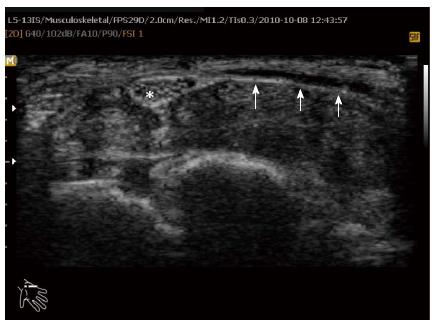
Figure 4 Axial ultrasound image shows flexor retinaculum bowing as an echogenic line (arrow) in carpal tunnel and cross sectional area of median nerve (stellate) in a patient with carpal tunnel syndrome.
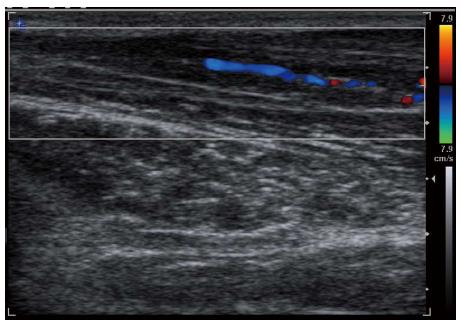
Figure 5 Longitudinal color Doppler sonogram in a 40-year-old woman with severe carpal tunnel syndrome shows intraneural hypervascularity in the median nerve.
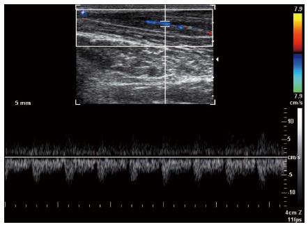
Figure 6 Spectral Doppler waveform of the median nerve shows low resistance hypervascularity of affected median nerve in a 40-year-old woman with severe carpal tunnel syndrome.
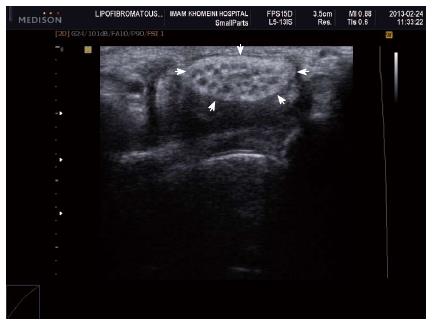
Figure 7 Axial ultrasound image shows hypoechoic cable like neural fascicle (arrows) separated by substratum hyperechoic fat in a patient with secondary carpal tunnel syndrome due to lipofibromatous hamartoma of the median nerve.
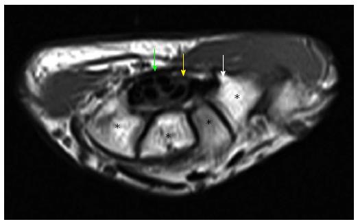
Figure 8 Axial T1W image of carpal tunnel at the level of tunnel outlet shows bony part of carpal tunnel as intermediate signal intensity composed from left to right hamate, capitates, trapezoid, trapezium.
White arrow shows hook of hamate, yellow arrow shows median nerve, green arrow shows flexor retinaculum. Asterisks indicate carpal bones.
- Citation: Ghasemi-rad M, Nosair E, Vegh A, Mohammadi A, Akkad A, Lesha E, Mohammadi MH, Sayed D, Davarian A, Maleki-Miyandoab T, Hasan A. A handy review of carpal tunnel syndrome: From anatomy to diagnosis and treatment. World J Radiol 2014; 6(6): 284-300
- URL: https://www.wjgnet.com/1949-8470/full/v6/i6/284.htm
- DOI: https://dx.doi.org/10.4329/wjr.v6.i6.284


