Copyright
©2013 Baishideng Publishing Group Co.
World J Radiol. Aug 28, 2013; 5(8): 325-327
Published online Aug 28, 2013. doi: 10.4329/wjr.v5.i8.325
Published online Aug 28, 2013. doi: 10.4329/wjr.v5.i8.325
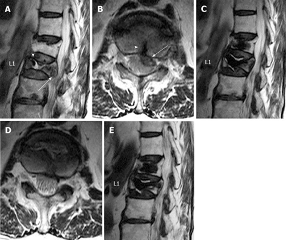
Figure 1 A 73-year-old woman with back pain (visual analog scale = 4 with medication).
A: A preoperative sagittal T2-weighted image shows unhealed compression fracture at L1 with an intravertebral cleft (arrowhead). A hypointense structure likely representing an epidural hematoma (EDH) is seen (arrow). There is a chronic compression fracture at T12; B: A preoperative axial T2-weighted image shows that a hypointense hematoma in the anterior aspect of the thecal sac at L1 (arrow). There is a fracture line (arrow head) extending into the EDH; C: A sagittal T2-weighted image obtained three months after the treatment shows the intervertebral cleft at L1 is filled with cement. Complete resolution of the EDH is noted. Complete pain relief was also noted at this time without medication (visual analog scale = 0); D: An axial T2-weighted image obtained three months after the treatment shows complete resolution of the EDH, resulting in amelioration of spinal canal compromise; E: A sagittal T2-weighted image obtained three years after the treatment shows maintenance of spinal canal without subsequent compression fracture.
- Citation: Hirata H, Hiwatashi A, Yoshiura T, Togao O, Yamashita K, Kamano H, Kikuchi K, Honda H. Resolution of epidural hematoma related to osteoporotic fracture after percutaneous vertebroplasty. World J Radiol 2013; 5(8): 325-327
- URL: https://www.wjgnet.com/1949-8470/full/v5/i8/325.htm
- DOI: https://dx.doi.org/10.4329/wjr.v5.i8.325


