Copyright
©2013 Baishideng Publishing Group Co.
World J Radiol. May 28, 2013; 5(5): 220-225
Published online May 28, 2013. doi: 10.4329/wjr.v5.i5.220
Published online May 28, 2013. doi: 10.4329/wjr.v5.i5.220
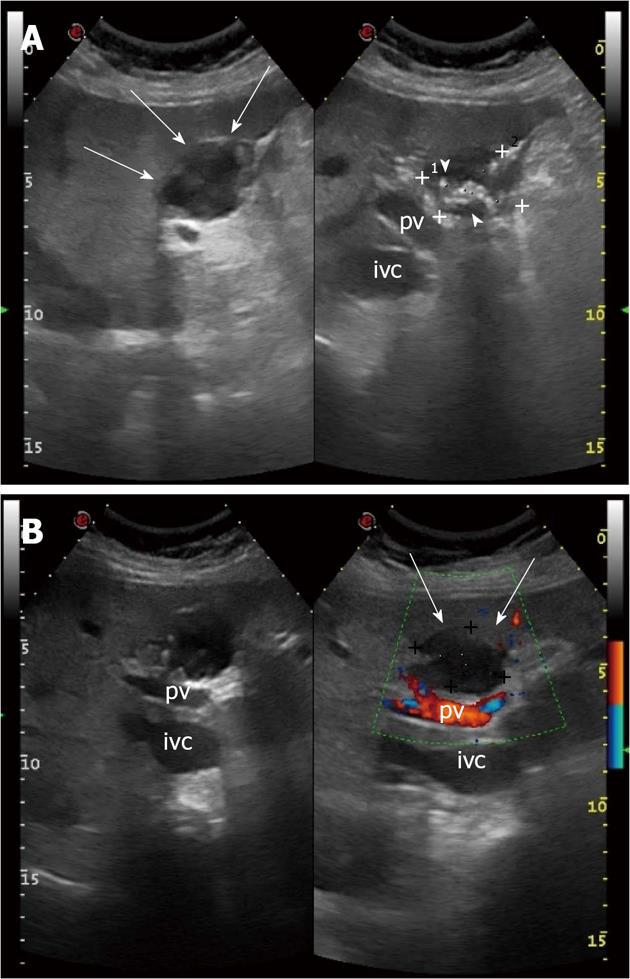
Figure 1 Chronic biloma in a 72-year-old woman: ultrasound findings.
A: Oblique view shows a heterogeneous hypo-anechoic rounded lesion with hyperechoic, calcified walls (arrows), numerous hyperechoic debris generating acustic shadow (arrow heads) and maximum size of 3.89 cm (caliper 1) × 3.42 cm (caliper 2); B: Color Doppler sonogram shows absence of vascularity inside the lesion (arrows). pv: Portal vein; ivc: Inferior vena cava.
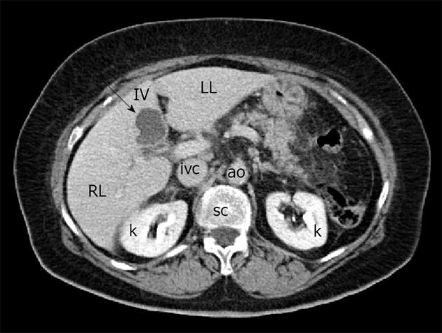
Figure 2 Computed tomography shows the biloma as a hypodense lesion in the IV hepatic segment, characterized by absence of enhancement after administration of intravenous contrast agent (arrow).
RL: Right hepatic lobe; LL: Left hepatic lobe; IV: Fourth hepatic segment; k: Kidney; ivc: Inferior vena cava; ao: Aorta; sc: Spinal column.
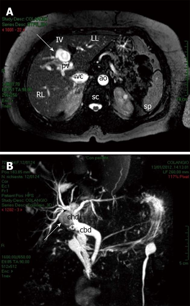
Figure 3 Biloma features on magnetic resonance imaging.
A: On T2-weighted images, the biloma appeared as a hyperintense lesion located in the IV hepatic segment (arrow); B: Contrast enhanced magnetic resonance cholangiopancreatography shows the biloma as a well-defined, rounded lesion (arrow) arising posteriorly to the confluence of right and left hepatic ducts into the common hepatic duct (arrowhead) in proximity to the stump of the remnant cystic duct (star). RL: Right hepatic lobe; LL: Left hepatic lobe; IV: Fourth hepatic segment; ivc: Inferior vena cava; ao: Aorta; pv: Portal vein; sc: Spinal column; sp: Spleen; chd: Common hepatic duct; cbd: Common bile duct.
- Citation: Tana C, D’Alessandro P, Tartaro A, Tana M, Mezzetti A, Schiavone C. Sonographic assessment of a suspected biloma: A case report and review of the literature. World J Radiol 2013; 5(5): 220-225
- URL: https://www.wjgnet.com/1949-8470/full/v5/i5/220.htm
- DOI: https://dx.doi.org/10.4329/wjr.v5.i5.220


