Copyright
©2012 Baishideng Publishing Group Co.
World J Radiol. May 28, 2012; 4(5): 231-235
Published online May 28, 2012. doi: 10.4329/wjr.v4.i5.231
Published online May 28, 2012. doi: 10.4329/wjr.v4.i5.231
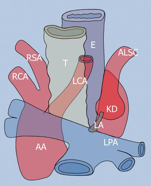
Figure 1 Schematic drawing of the right-sided aortic arch with aberrant left subclavian artery and left ligamentum arteriosum.
The left ligamentum arteriosum proximal attachment is represented by Kommerell’s diverticulum (KD) which is the broad base from which the aberrant left subclavian artery originates. AA: Ascending aorta; LPA: Left pulmonary artery; LA: Ligamentum arteriosum; LCA: Left common carotid artery; RSA: Right subclavian artery; RCA: Right common carotid artery; T: Trachea; E: Esophagus.
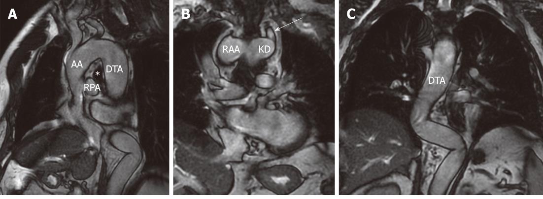
Figure 2 Cardiac-gated FIESTA images.
A: Sagittal oblique cardiac-gated FIESTA image (A) demonstrating the right aortic arch above the right main bronchus (asterisk) and right main pulmonary artery; B, C: Coronal cardiac-gated FIESTA images showing the relationships between the RAA and Kommerell's diverticulum from which the ALSC (large arrow in B) originates. The descending thoracic aorta courses on the right side of the spine and eventually turns on the left side to enter the aortic diaphragmatic hiatus. Large arrow indicate aberrant left subclavian artery. AA: Ascending aorta; DTA: Descending thoracic aorta; *: Right main bronchus; RPA: Right pulmonary artery; RAA: Right aortic arch; KD: Kommerell’s diverticulum.
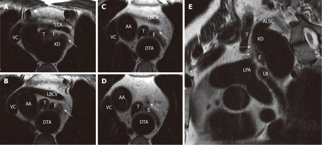
Figure 3 Four sequential frFSE T2-weighted cardiac-gated axial images (A-D) and sequence (E).
A-D: The ligamentum arteriosum from its proximal attachment on Kommerell’s diverticulum (KD) to its distal anchorage site on the superior aspect of the left pulmonary artery (LPA) origin; E: The complete course of the ligamentum arteriosum, which is appreciable anteriorly to the esophagus and trachea. Arrows indicate ligamentum arteriosum. VC: Vena cava; LBCV: Left brachio-cephalic vein; T: Trachea; E: Esophagus; LCA: Left common carotid artery; AA: Ascending aorta; DTA: Descending thoracic aorta; ALSC: Aberrant left subclavian artery.
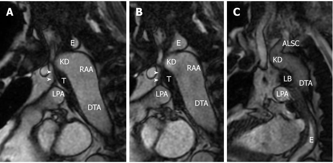
Figure 4 Three sequential frames of a cine-FIESTA sagittal oblique sequence.
A, B: Progressive distension of the esophageal lumen superiorly to the right aortic arch (RAA); C: The water bolus is appreciable in the lower thoracic esophagus, but it was impossible to obtain a significant distention of the esophageal segment passing through the vascular ring formed in its left lateral inferior part by the ligamentum arteriosum. Arrowheads indicate ligamentum arteriosum. T: Trachea; E: Esophagus; ALSC: Aberrant left subclavian artery; KD: Kommerell’s diverticulum; DTA: Descending thoracic aorta; LPA: Left pulmonary artery.
- Citation: Paparo F, Bacigalupo L, Melani E, Rollandi GA, De Caro G. Cardiac-MRI demonstration of the ligamentum arteriosum in a case of right aortic arch with aberrant left subclavian artery. World J Radiol 2012; 4(5): 231-235
- URL: https://www.wjgnet.com/1949-8470/full/v4/i5/231.htm
- DOI: https://dx.doi.org/10.4329/wjr.v4.i5.231


