Copyright
©2012 Baishideng Publishing Group Co.
World J Radiol. May 28, 2012; 4(5): 231-235
Published online May 28, 2012. doi: 10.4329/wjr.v4.i5.231
Published online May 28, 2012. doi: 10.4329/wjr.v4.i5.231
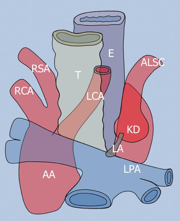
Figure 1 Schematic drawing of the right-sided aortic arch with aberrant left subclavian artery and left ligamentum arteriosum.
The left ligamentum arteriosum proximal attachment is represented by Kommerell’s diverticulum (KD) which is the broad base from which the aberrant left subclavian artery originates. AA: Ascending aorta; LPA: Left pulmonary artery; LA: Ligamentum arteriosum; LCA: Left common carotid artery; RSA: Right subclavian artery; RCA: Right common carotid artery; T: Trachea; E: Esophagus.
- Citation: Paparo F, Bacigalupo L, Melani E, Rollandi GA, De Caro G. Cardiac-MRI demonstration of the ligamentum arteriosum in a case of right aortic arch with aberrant left subclavian artery. World J Radiol 2012; 4(5): 231-235
- URL: https://www.wjgnet.com/1949-8470/full/v4/i5/231.htm
- DOI: https://dx.doi.org/10.4329/wjr.v4.i5.231


