Copyright
©2012 Baishideng Publishing Group Co.
World J Radiol. May 28, 2012; 4(5): 215-219
Published online May 28, 2012. doi: 10.4329/wjr.v4.i5.215
Published online May 28, 2012. doi: 10.4329/wjr.v4.i5.215
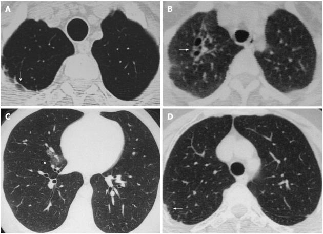
Figure 1 High resolution computed tomography images.
A: Subpleural bands (arrow); B: Apical fibrosis and bronchial dilatation (arrow); C: Ground-glass opacity (arrow); D: Irregularity of interfaces (arrow).
- Citation: Hasiloglu ZI, Havan N, Rezvani A, Sariyildiz MA, Erdemli HE, Karacan I. Lung parenchymal changes in patients with ankylosing spondylitis. World J Radiol 2012; 4(5): 215-219
- URL: https://www.wjgnet.com/1949-8470/full/v4/i5/215.htm
- DOI: https://dx.doi.org/10.4329/wjr.v4.i5.215


