Copyright
©2012 Baishideng Publishing Group Co.
World J Radiol. Nov 28, 2012; 4(11): 450-454
Published online Nov 28, 2012. doi: 10.4329/wjr.v4.i11.450
Published online Nov 28, 2012. doi: 10.4329/wjr.v4.i11.450
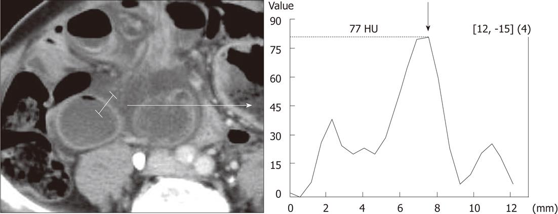
Figure 1 Measurement method of the computed tomography value of the intestinal tract wall.
The computed tomography (CT) value was continuously measured from the lumen of the enlarged intestinal tract toward the outer side, with the highest value set as the CT value of the intestinal tract wall.
Figure 2 Example: Male patient aged 70 yr.
The patient visited the emergency room due to abdominal pain at night and was hospitalized. The patient was diagnosed with strangulation ileus, resulting in emergency surgery, and an enterectomy was performed. On the computed tomography image at the time of hospitalization, the poor contrast effect is easily observed for the ischemic bowel wall (arrows) compared to non-ischemic bowel wall in the arterial phase (A), however, it is not observed in the portal vein phase (B) and the equilibrium phase (C).
- Citation: Ohira G, Shuto K, Kono T, Tohma T, Gunji H, Narushima K, Imanishi S, Fujishiro T, Tochigi T, Hanaoka T, Miyauchi H, Hanari N, Matsubara H, Yanagawa N. Utility of arterial phase of dynamic CT for detection of intestinal ischemia associated with strangulation ileus. World J Radiol 2012; 4(11): 450-454
- URL: https://www.wjgnet.com/1949-8470/full/v4/i11/450.htm
- DOI: https://dx.doi.org/10.4329/wjr.v4.i11.450


