Copyright
©2011 Baishideng Publishing Group Co.
World J Radiol. Jul 28, 2011; 3(7): 194-198
Published online Jul 28, 2011. doi: 10.4329/wjr.v3.i7.194
Published online Jul 28, 2011. doi: 10.4329/wjr.v3.i7.194
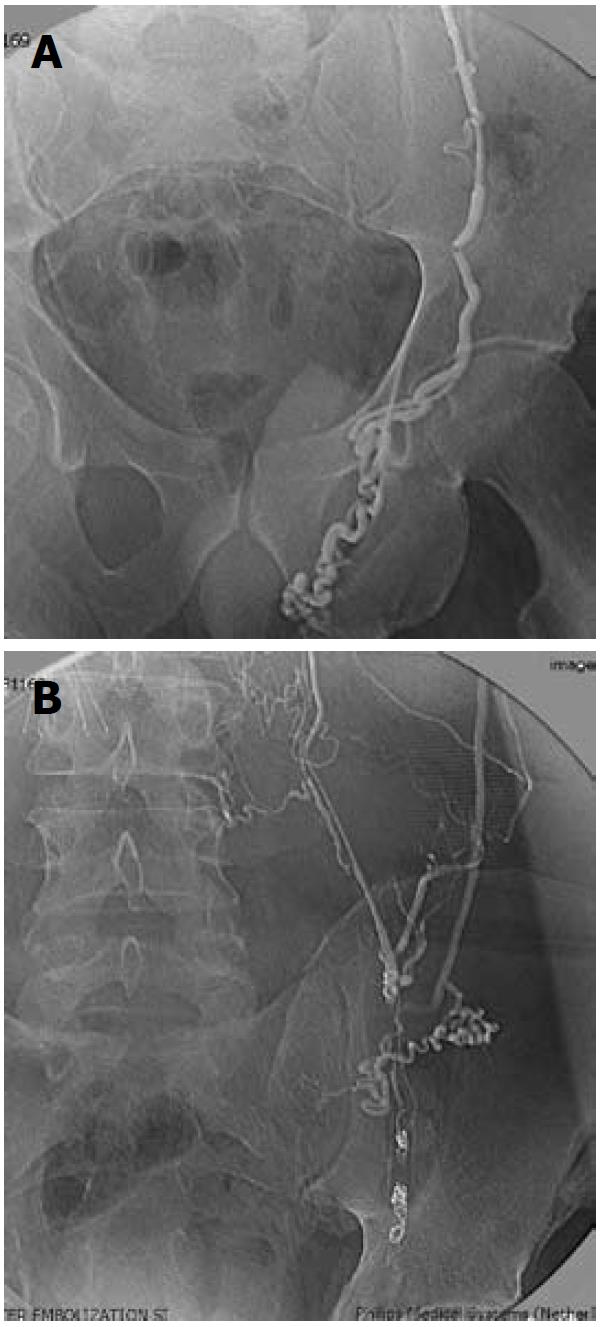
Figure 1 Venography following transjugular access.
A: Left gonadal venogram with transjugular access demonstrating large left varicocele; B: Left gonadal venogram, following coil occlusion of the gonadal vein at the internal inguinal ring, demonstrating extensive retroperitoneal collateralization.
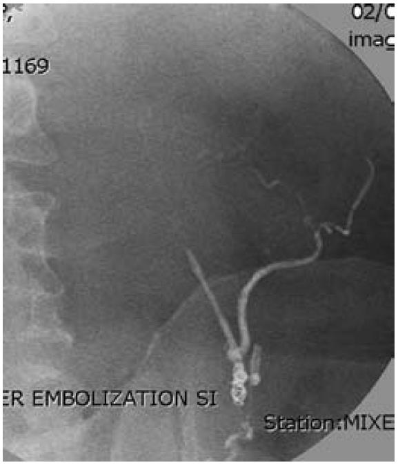
Figure 2 Repeat venography via transjugular access, showing a cyanoacrylate cast of left gonadal vein and collaterals following coil placement and cyanoacrylate occlusion of collateral circulation.
Figure 3 Direct percutaneous pampiniform venography performed under ultrasound guidance.
A: Collateral flow into the saphenous (arrow) and femoral (arrowhead) venous system through the external pudendal vein (double arrowhead). Cross filling from right to left through the scrotal veins is also demonstrated (double arrow); B: Collateral flow to the saphenous vein (arrow) through the external pudendal vein (double arrowhead) and to the external iliac vein through the cremasteric/inferior epigastric vein (double arrow).
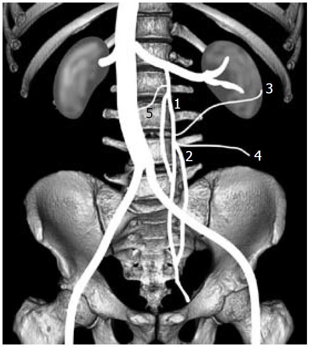
Figure 4 Diagram illustrating retroperitoneal collaterals to the left gonadal vein.
Gonadal vein (1) parallel retroperitoneal vein (2) renal capsular collateral vein (3) axial lateral retroperitoneal collateral vein to the colon (4) axial medial collateral (5).
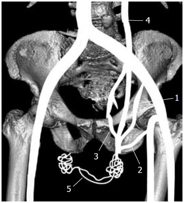
Figure 5 Diagram illustrating trans-scrotal collateral communication with the pampiniform plexus.
Cremasteric (external spermatic) vein (1) to inferior epigastric/external iliac vein; external pudendal vein (2) to saphenous vein; vein of the vas deferens (3) to internal iliac vein; left gonadal vein (4) trans-scrotal collateral veins (5).
- Citation: Gendel V, Haddadin I, Nosher JL. Antegrade pampiniform plexus venography in recurrent varicocele: Case report and anatomy review. World J Radiol 2011; 3(7): 194-198
- URL: https://www.wjgnet.com/1949-8470/full/v3/i7/194.htm
- DOI: https://dx.doi.org/10.4329/wjr.v3.i7.194


